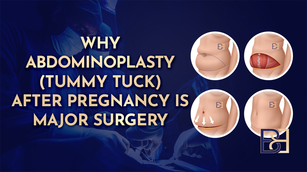
This is a step by step account of one body contouring operation I performed for a post pregnancy patient. It is written to show what actually happens during surgery, stage by stage, using photographs taken during the case. Every patient is different, and this account describes one person’s operation, not a template for what yours would involve.
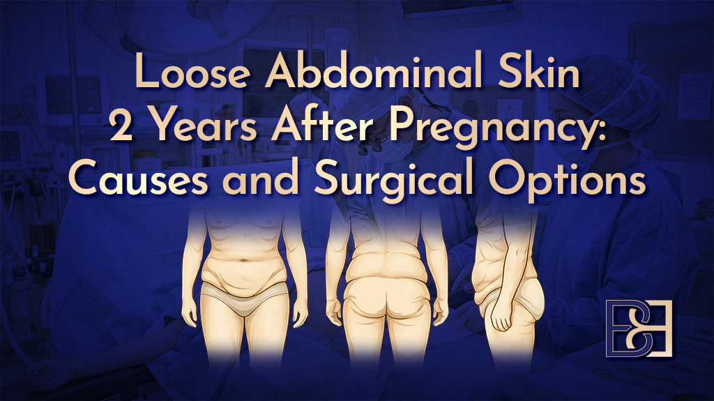
The patient was a 44 year old woman who had completed her family. After her pregnancies she had returned to her usual weight. She presented with a moderate amount of loose abdominal skin that extended past the lower abdomen towards the hips, separation of the abdominal muscles (diastasis recti), and a left femoral hernia that was causing her pain and discomfort.
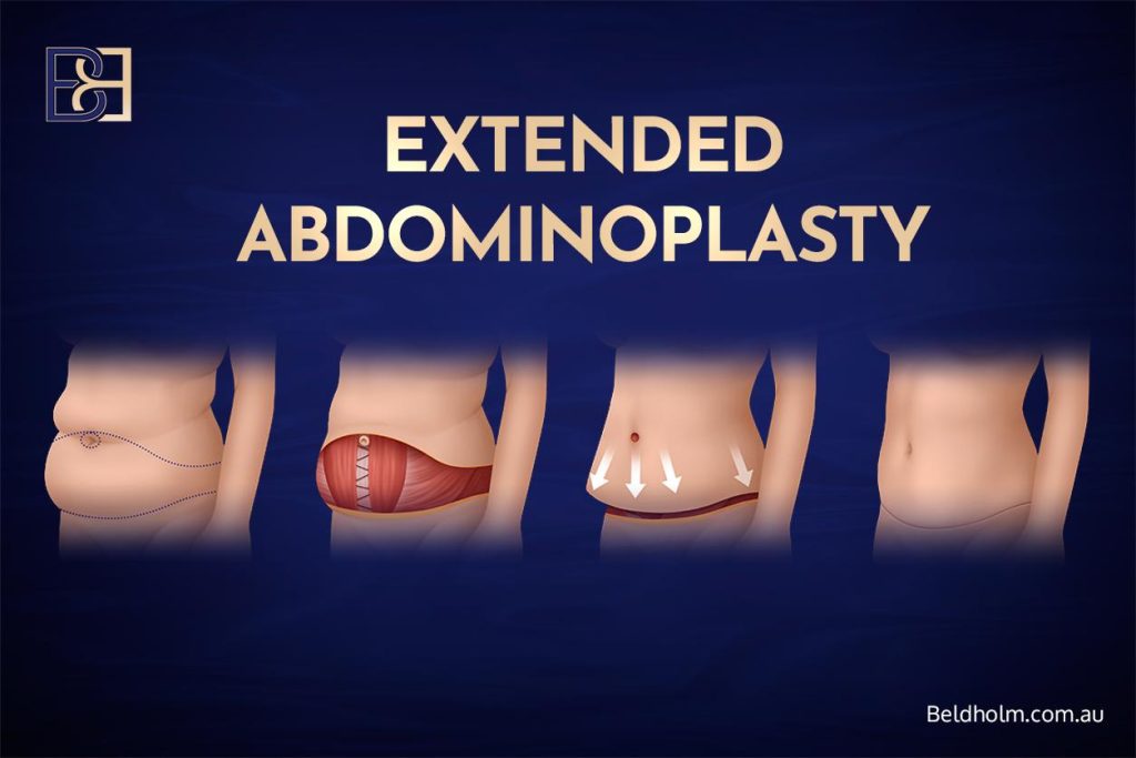
Because the femoral hernia was symptomatic and sat within the same area I was already operating on, I repaired it through the same incision during the one operation, rather than asking her to return for a separate procedure and a second anaesthetic. The surgery I performed was an extended abdominoplasty (tummy tuck) with ultrasound-assisted suction lipectomy (VASER liposuction), diastasis recti repair, and left femoral hernia repair.
Preparing the patient and marking up in the anaesthetic bay
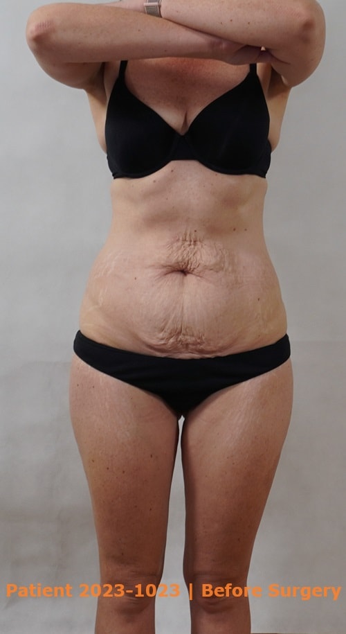
On the day of surgery I met the patient in the anaesthetic bay. I went over her medical history again, answered her remaining questions, and confirmed the plan for the operation with her.
I then marked her up while she was standing upright. Marking a patient standing is important, because loose skin and the abdominal contour sit differently when a person is lying flat on the operating table. Standing markings let me plan the incision line, the extent of skin to be removed, and the areas for the ultrasound-assisted suction lipectomy (VASER liposuction) accurately before she was anaesthetised.
Once the markings were complete, the anaesthetist gave her premedication. She was then taken into the operating theatre and given a general anaesthetic for the procedure.
The initial abdominoplasty incision
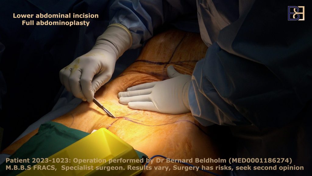
The abdominoplasty procedure began with a low, horizontal incision across the lower abdomen. In this patient the incision continued past the hip bones on each side, which is what makes it an extended abdominoplasty (extended tummy tuck) rather than a standard full abdominoplasty.
The difference comes down to how far the loose skin reaches. In a full or traditional abdominoplasty (tummy tuck) the incision runs from one hip bone to the other. When loose skin carries on around towards the lateral hip, as it did here, the incision is carried further out past the hip bone so that skin can be removed as well. In post pregnancy patients this lateral extension is usually modest, and that was the case for this patient.
At this stage I also released the umbilicus (belly button) from the surrounding skin, leaving it attached to its stalk on the abdominal wall. This lets the skin above be lifted and redraped later while the umbilicus stays in its correct anatomical position.
Repairing the femoral hernia through the same incision
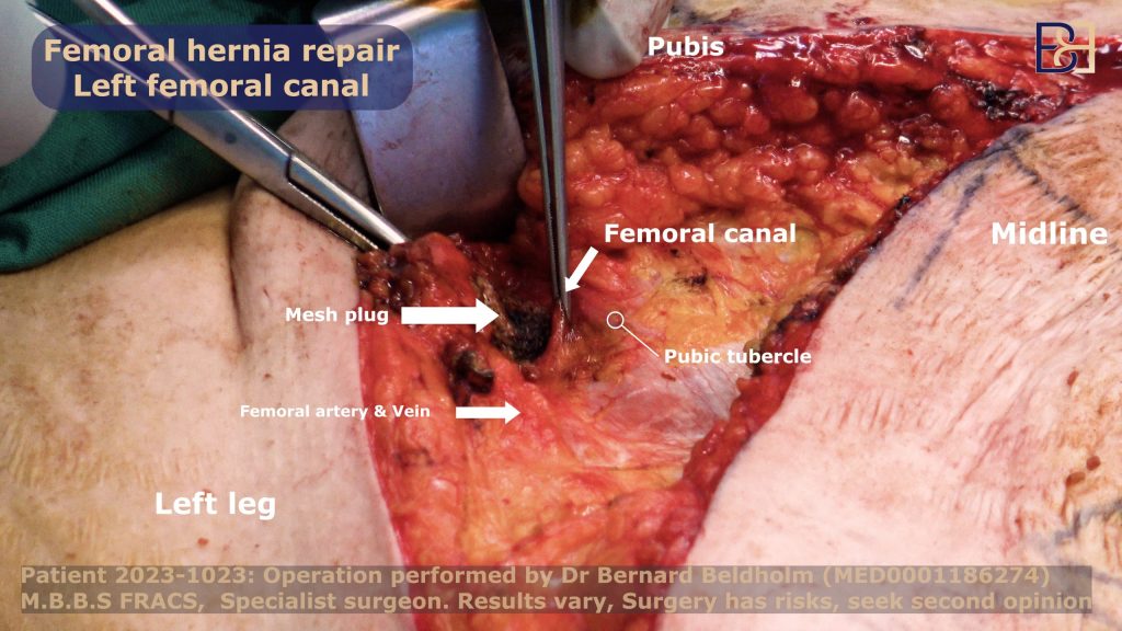
With the lower abdomen open, I moved to the left femoral hernia. A femoral hernia occurs when tissue pushes through a weak point in the groin, below the inguinal ligament. Femoral hernias are more common in women, and this one was symptomatic, causing the patient pain and discomfort.
A femoral hernia is usually repaired on its own through a small, separate incision in the groin. In this case the hernia sat within the field I had already opened for the abdominoplasty, so I repaired it through the same incision. That meant one operation and one anaesthetic for both problems, rather than a second procedure on another day.
To repair the hernia I reduced the herniated tissue back into place and reinforced the weak point with mesh, held with permanent sutures. The mesh becomes incorporated as the tissue heals around it, giving a durable repair.
Ultrasound-assisted suction lipectomy (VASER liposuction)
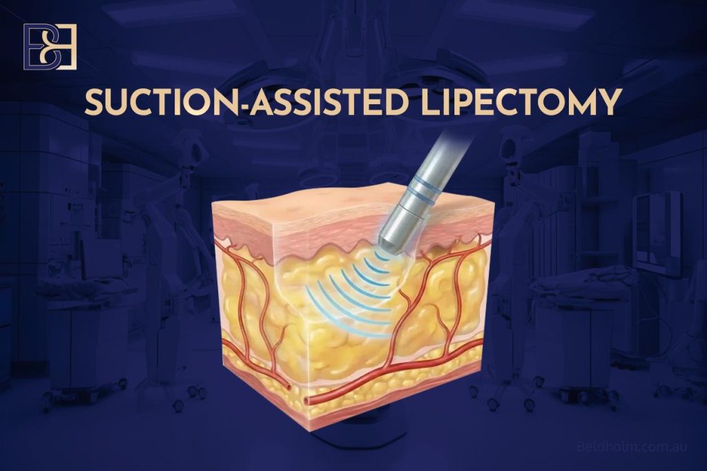
This patient carried very little excess fat, so the ultrasound-assisted suction lipectomy (VASER liposuction) part of the operation was brief, around 10 to 20 minutes, focused on the flanks and the mons pubis (pubic area). The steps are the same regardless of how much is treated.
- I first inject tumescent fluid, a mix of saline with adrenaline and local anaesthetic, into the target area. This reduces bleeding and helps separate the fat.
- I make a small entry incision and place protective ports in the skin. The VASER probe generates heat, and the ports protect the skin edges from thermal injury.
- I pass the ultrasound probe through the port. When it is switched on, the ultrasound energy breaks down the fat.
- Once the fat has been emulsified, I remove the probe and insert a cannula, a thin hollow tube, connected to suction.
- The suction removes the treated fat.
Treating the fat this way, rather than by wide surgical dissection, means I disturb less of the surrounding tissues in the areas that are suctioned.
Handling the umbilicus (belly button)
The umbilicus is tethered by a stalk to the abdominal wall, and it sits in a fixed anatomical position on the muscle layer. The skin around it, though, is part of the sheet of skin that gets lifted and redraped during an abdominoplasty.
If the umbilicus were left attached to the surrounding skin, it would be dragged downward as that skin is pulled taut, ending up out of position. To avoid this, I cut around the umbilicus and free it from the skin while leaving it connected to its stalk. It stays in place on the abdominal wall while the skin is redraped over it. Later in the operation I bring it out through a new opening made in the redraped skin, so it sits where it should.
Lifting the abdominal skin flap
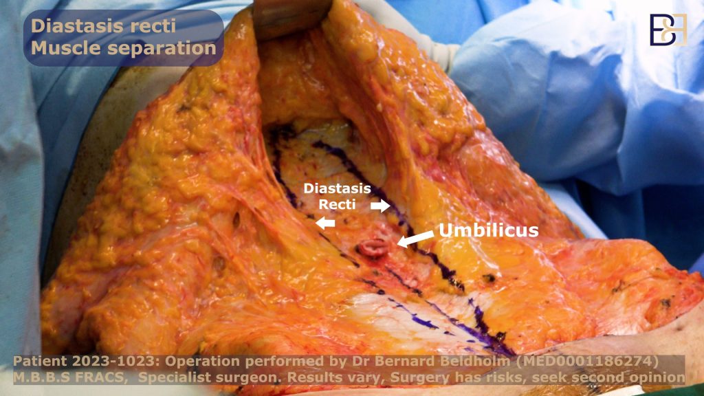
Next I lifted the skin off the underlying muscles and abdominal wall. I make my incisions straight, at 90 degrees to the skin, rather than at an angle, because straight edges tend to heal more neatly.
As I lift the flap, I stay just below Scarpa’s fascia, a layer of connective tissue in the abdominal wall. Working at this level preserves the blood vessels that run in the flap, which matters for wound healing. If a small vessel bleeds during the dissection, I seal it with diathermy, which uses heat to coagulate the tissue and stop the bleeding.
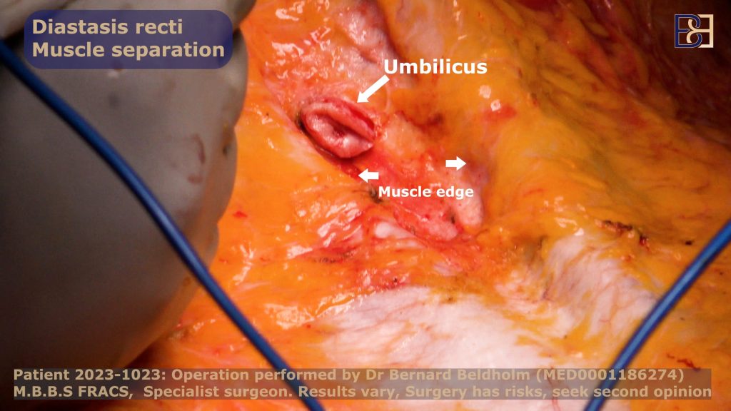
Once I reach the level of the umbilicus, I make a narrow tunnel up towards the ribs, over the upper abdomen. This gives me a clear view of the linea alba, the strip of connective tissue running down the midline, so I can see the full extent of the muscle separation before repairing it.
Repairing the diastasis recti (muscle separation)
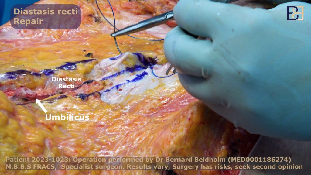
The two rectus abdominis muscles run vertically down the front of the abdomen and are joined at the midline by a strip of connective tissue called the linea alba. During pregnancy the growing uterus stretches the linea alba, and the muscles move apart. This separation is called diastasis recti. It is a normal change after pregnancy, and in many women some degree of it remains once the pregnancies are over, particularly after multiple pregnancies (1).
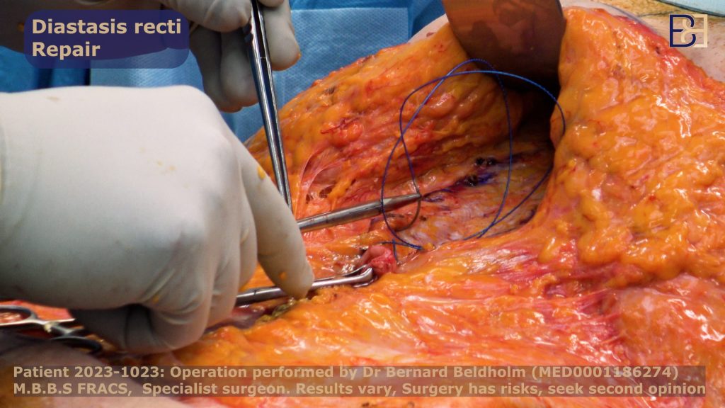
Once I had exposed the midline, I could see the extent of the separation in this patient. To bring the separated abdominal muscles back together, I stitch the two sides of the linea alba towards the midline, closing the gap. I use barbed sutures (V-Loc), which hold tension along their length and do not need to be knotted.
I do this diastasis repair in two layers. The deeper layer takes the tension of bringing the muscles together. The final layer sits only in the rectus sheath, the tough covering over the muscle, and does not go any deeper than that, so I stay clear of the nerves, muscle and blood vessels beneath it.
Closing the dead space and finishing without drains
When the skin flap is laid back down over the abdominal wall, a potential space is left between the two. Fluid can collect in that space and form a seroma. To manage this, I use progressive tension sutures, which tack the flap down to the abdominal wall in rows as I advance towards the incision.
These sutures do two things. They close down the space between the flap and the wall, which lowers the chance of a seroma, and they take the tension off the final skin incision by holding it across the abdominal wall instead. That means the wound is not carrying the load on its own. I also apply tissue glue, which further reduces the space where fluid can gather.
Because this technique manages the dead space directly, I was able to close this patient without surgical drains. Most of my post pregnancy abdominoplasty patients are closed drainless in the same way. Drains are reserved for cases with a larger amount of loose skin.
Marking and removing the excess skin
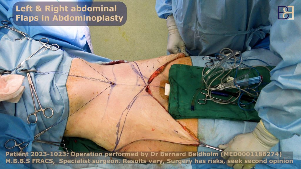
With the skin redraped and held at tension, and before I place the final sutures, I remove the skin that is surplus to a smooth closure.
Some surgeons trim the excess in segments as they close. I prefer to mark the full amount to be removed first. I lift the redraped skin with Alice forceps along the intended line, check that the amount is correct, and then resect the skin in one go along that mark. Marking the whole resection before cutting gives me an even, symmetrical result and avoids removing too much.
For this patient the excess tissue removed weighed 337 grams on the right side and 382 grams on the left.
Redraping the skin and setting the umbilicus
With the muscle repair done, there was a sheet of loose skin to redrape and trim. To judge how much skin to remove and how much tension to set, I flex the operating table, tilting it about 15 to 20 degrees so the patient is jack-knifed at the hips. This brings the upper and lower skin edges together the way they will sit once she is upright, so I am not setting the skin too tight.
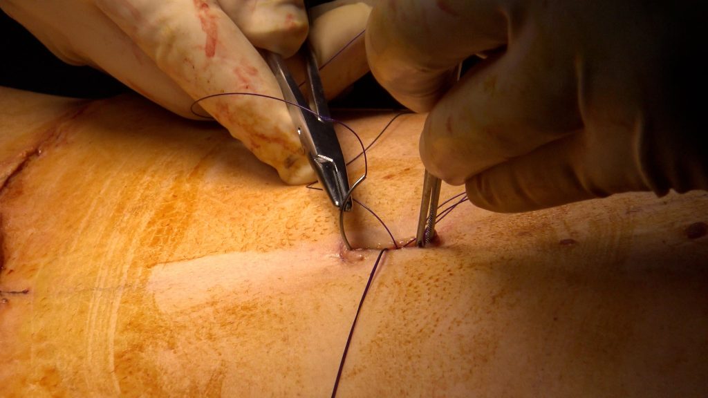
With the skin held at the right tension, I turn to the umbilicus. I find its position under the redraped skin, make a new opening, and bring the stalk through. I trim any excess fat around it so it sits flush, then stitch it into place in its new opening.
Dressing and compression garment
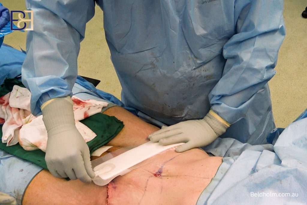
Once the wound was closed, I applied a PICO dressing over the incision. PICO is a single-use negative pressure wound therapy system. A small battery-powered pump applies low-level suction across the wound, holding the edges together, drawing off excess fluid, and keeping the area clean while it heals. The dressing runs at a nominal 80 mmHg and stays on for about 7 days.
I use PICO on the incision for most of my abdominoplasty patients because managing wound fluid and keeping the edges supported in the first week helps healing and lowers the chance of wound problems.
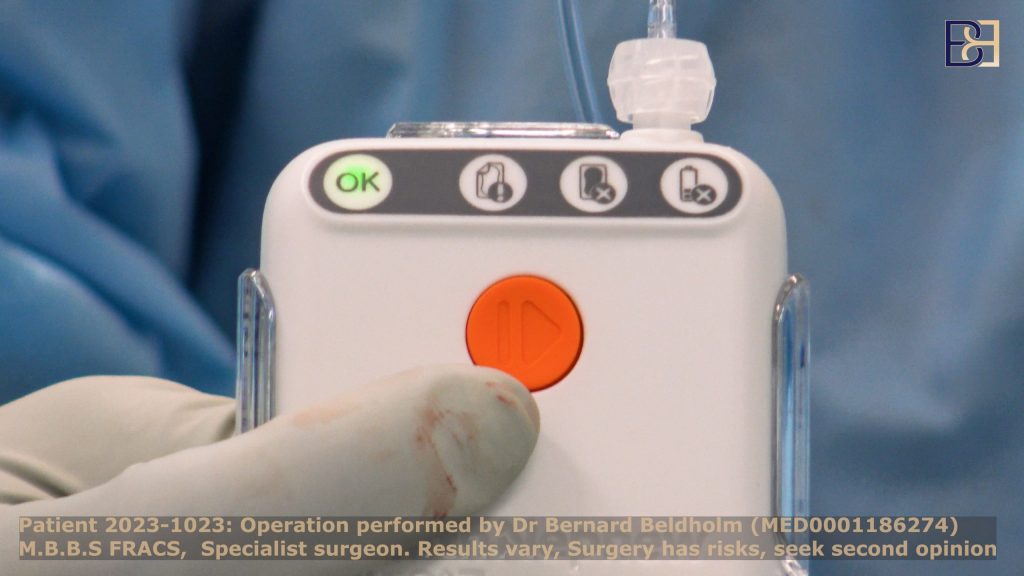
Over the top, the patient wears a compression garment. The garment supports the abdominal area, helps control swelling, and is part of the recovery routine for the weeks after surgery. I will go through how long to wear it, and what to expect week to week, at the follow-up appointments.
Book your appointment online now
What was done in this operation
For reference, here is the operative record for this patient in one place.
- Extended abdominoplasty (tummy tuck), incision carried past the hip bones
- Ultrasound-assisted suction lipectomy (VASER liposuction): 100 ml of fat removed from the flanks and mons pubis
- Excess abdominal skin removed: 337 grams on the right, 382 grams on the left
- Diastasis recti (muscle separation) repaired
- Left femoral hernia repaired with mesh
- Closed without drains, PICO dressing applied
Risks and recovery realities
This is major surgery. Combining an extended abdominoplasty, ultrasound-assisted suction lipectomy (VASER liposuction), diastasis recti repair and a hernia repair in one operation means a longer time under anaesthetic and a bigger physiological event than any one of those steps alone. That has to be weighed up honestly before deciding on surgery.
Risks
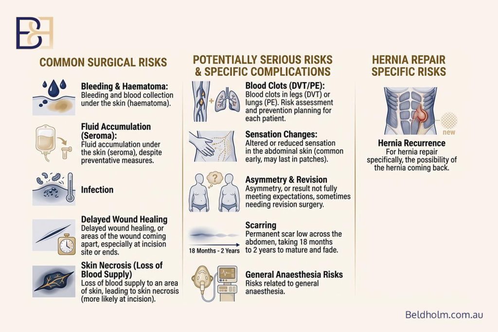
Every surgical procedure carries risk. For this type of surgery the ones I discuss with patients include:
- Bleeding and blood collection under the skin (haematoma)
- Fluid accumulation under the skin (seroma), even with the measures used to reduce it
- Infection
- Delayed wound healing, or areas of the wound coming apart, which can happen more at the incision site, at the ends of the wound
- Loss of blood supply to an area of skin, which can lead to skin necrosis, and is more likely at the incision site
- Blood clots in the legs or lungs (deep vein thrombosis and pulmonary embolism). I assess each patient’s clot risk myself and plan their prevention accordingly
- Altered or reduced sensation in the abdominal skin, which is common early on and can be long lasting in patches
- Asymmetry, or a result that does not fully meet what was hoped for, which can sometimes need revision surgery
- Scarring. The scar is permanent, sits low across the abdomen, and takes 18 months to 2 years to mature and fade
- Risks related to the general anaesthesia
- For the hernia repair specifically, the possibility of the hernia coming back, and risks related to the mesh
This is not a complete list, and I go through the risks relevant to each patient at consultation.
Recovery
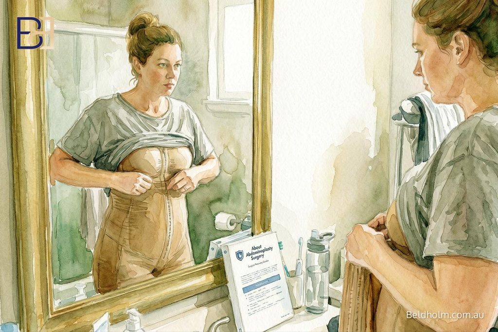
Most patients stay in hospital for 1 to 2 days. The muscle repair in particular makes the first couple of weeks uncomfortable, because the tightened abdominal wall is under tension. Pain is managed, but it is real.
A few practical points I make clear before surgery:
- You will be moving slowly and cannot do any heavy lifting for several weeks
- A compression garment is worn for weeks after the operation
- You will need help at home. This matters a lot for post pregnancy patients, because caring for young children involves exactly the lifting, bending and carrying that you need to avoid early on. Planning that support in advance is important
- Returning to exercise and normal activity is staged over weeks to months, not days. Strenuous exercise and strenuous activity wait until later in recovery
- Rebuilding your core muscles is staged and guided, because the muscle repair needs time to hold
Results differ from one patient to the next, depending on anatomy, healing and the extent of surgery. I review every patient in person at 4 weeks, 3 months, 6 months and 12 months to follow their healing.
Timing surgery after pregnancy and breastfeeding

I plan this post pregnancy surgery once a patient has finished having children and has finished breastfeeding. There are good reasons for both.
If you go on to have another pregnancy after an abdominoplasty, it can stretch the skin and separate the repaired muscles again, which can undo the result. For that reason I recommend surgery once your family is complete.
Breastfeeding is a separate consideration. It affects the breasts rather than the abdominal wall, but I still wait until it has finished and the breasts and the body have settled after the most recent pregnancy before planning surgery.
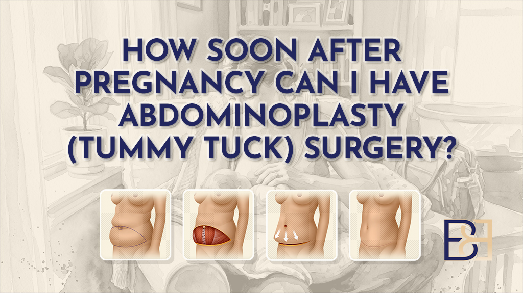
As a general guide this often works out to somewhere around 12 months after the most recent pregnancy, but that is not a fixed rule. The right timing depends on the individual and is decided at consultation, not set by a number on a page.
Frequently asked questions
What is diastasis recti?
Diastasis recti is separation of the two rectus abdominis muscles at the midline, where the connective tissue between them (the linea alba) has stretched. It is a normal change during and after pregnancy, as the growing uterus stretches the abdominal wall. In many women some separation remains after the pregnancies are over. Whether you have it, and how much, is assessed at consultation.
How can I check for it at home?
There is a rough way to get a sense of it. Lie on your back with your knees bent and feet flat. Place your fingers along the midline of your abdomen, just above and just below the belly button. Lift your head and shoulders slightly off the floor, as if starting a sit-up. If you can feel a gap between the muscle edges, that may be diastasis recti. This is only a guide, not a diagnosis, and a proper assessment is done in person.
Is this surgery covered by Medicare?
Sometimes. Abdominoplasty (tummy tuck) with diastasis recti repair can attract a Medicare rebate under MBS item 30175 when specific eligibility criteria are met following pregnancy. Where the item applies, Medicare pays a benefit of 75% of the schedule fee, and a private health fund covers a further portion if you have appropriate cover. Where surgery does not meet the criteria and is cosmetic, no Medicare benefit applies and GST is added.
I do not publish dollar figures, because the MBS schedule fee is updated each year and out-of-pocket costs depend on your own cover. My patient coordinators obtain written quotes for the hospital and anaesthetist, and the practice provides written financial consent before surgery. For the current schedule fee, the MBS Online website has the live figures.
Can the hernia be repaired at the same time as the abdominoplasty?
In this patient, yes, because the femoral hernia was symptomatic and sat within the area already opened for the abdominoplasty. That let me repair both in one operation. Whether concurrent repair suits another patient depends on the type and position of the hernia, and is decided case by case.
References
- Soto-Gonzalez M, Da Cuna-Carrera I, Lantaron-Caeiro EM, Pascoal AG. Effect of hypopressive and conventional abdominal exercises on postpartum diastasis recti: a randomized controlled trial. PLoS One. 2024;19(12):e0314274.