


Mastopexy with auto augmentation is a breast lift (mastopexy) that adds volume to the upper part of the breast using your own tissue rather than an implant. During the operation I raise a flap of your own breast tissue and reposition it higher on the chest wall, so the upper pole carries more fullness while the nipple and areola are moved to a higher position. No device is placed. The volume comes from tissue you already have, redistributed and secured.
Because it combines a lift with a change in breast shape, it is a more involved operation than a lift on its own. As with any cosmetic surgery, it is a genuine physiological event, and it carries a defined set of risks. On this page I set those risks out directly so you can weigh them before deciding anything. My aim is not to talk you into or out of the operation. It is to give you an accurate picture of what can go wrong, how often, and what I do about it.
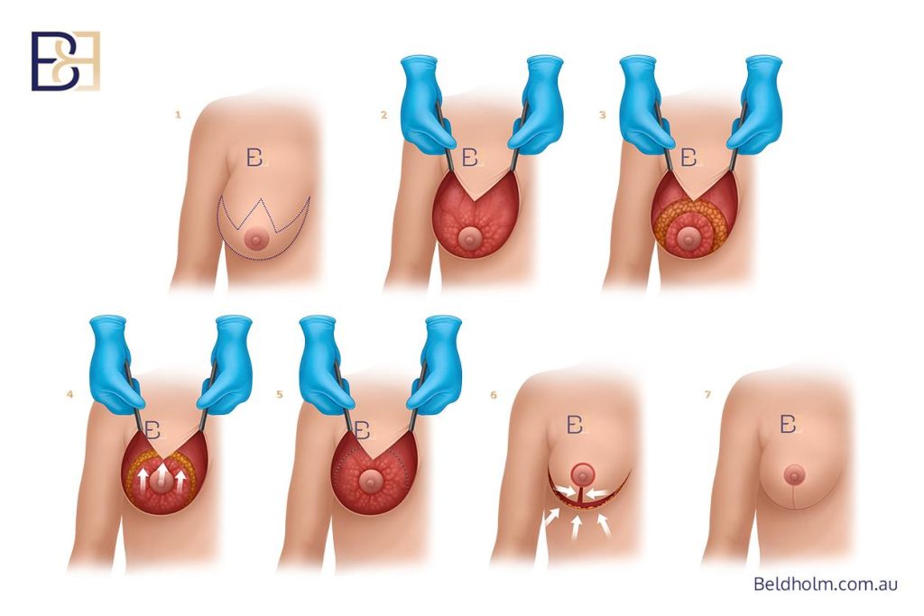
A few points apply throughout.
- Every patient heals differently, and results vary from person to person. Nothing here is a prediction of your individual outcome.
- A GP referral is required before any consultation.
This page covers complications only. Recovery timelines, activity restrictions and aftercare are covered separately.
What kind of risks this operation carries
The volume in this operation comes from your own tissue rather than an implant. Because no device is placed, the risks are those of a tissue-based breast operation, and they centre on a few things.
- The blood supply to the tissue that is moved, and to the nipple and areola
- Wound healing along the incisions
- How the flap behaves over time, including how well the added upper pole volume holds
- The scars, which are more extensive than a lift on its own
These are the areas the rest of this page works through, one at a time. Auto augmentation and breast implant augmentation are different operations with different risk profiles, and which one suits a given patient is a separate discussion for consultation. This page covers the risks of the auto augmentation operation specifically.
Bleeding and haematoma

Any breast operation involves cutting through tissue that has a blood supply, so some bleeding is expected and is controlled during surgery. The complication to watch for is a haematoma, which is a collection of blood that builds up under the skin or within the breast after the operation, usually in the first day or two.
A haematoma tends to show itself as one breast becoming firmer, more swollen, more painful, or more bruised than the other over a short period. A small haematoma may settle on its own and be managed by observation. A larger one usually needs a return to theatre to drain the blood and stop the bleeding point.
Across the published series on auto augmentation, haematoma is uncommon. In a series of just under 200 patients undergoing a mushroom-flap auto augmentation, one patient (around half of one per cent) developed a haematoma that needed a further operation (1). In an inferior-pedicle auto augmentation series, two patients (around four per cent) had a haematoma, one of which was drained on the day of surgery and healed without further trouble (2). In a dermal-suspension auto augmentation series, one haematoma required operative drainage (3).
Two things raise the chance of bleeding after surgery:
- Blood-thinning medication and some supplements. Aspirin and anticoagulants are usually stopped about a week before surgery, but only on a plan agreed in advance. You should never stop or adjust prescribed blood-thinning medication on your own.
- Raised blood pressure and physical strain in the early days after surgery.
I manage bleeding risk by controlling every bleeding point during the operation, and by reviewing your medications and health before surgery. If a haematoma does occur, it is treated promptly. Dealt with early, it usually does not affect the final result.
Infection
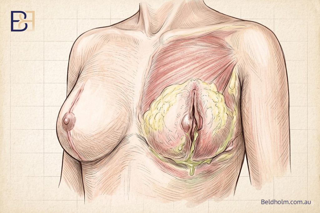
Any surgical wound can become infected. In breast surgery the risk is low, but it is not zero. Here is what an infection looks like and how it is managed.
A wound infection usually appears in the first week or two. The signs are increasing redness spreading out from the incision, warmth, swelling, increasing pain rather than settling pain, and sometimes fluid or pus draining from the wound. A fever can accompany it.
Most infections are dealt with by antibiotics, and settle without any further surgery. In the published auto augmentation series, this is the usual picture. In a mushroom-flap series of just under 200 patients, six (around three per cent) needed antibiotics for a wound infection, and none of these needed a surgical drainage procedure (1). Deeper or more established infections occasionally need the wound to be opened and washed out, but this is uncommon in this operation.
Infection matters more in a tissue-based operation than the raw numbers suggest, because the tissue being repositioned depends on a good blood supply to heal. An infection can interfere with healing at the incision lines and, less often, affect the moved tissue itself.
I reduce infection risk with antibiotics given around the time of surgery, sterile operating conditions, and careful wound closure. Not smoking around the time of surgery makes a measurable difference to wound healing and infection risk, which is why smoking cessation is part of the preparation. If an infection does develop, early treatment gives the best chance of resolving the infection.
Wound healing and dehiscence
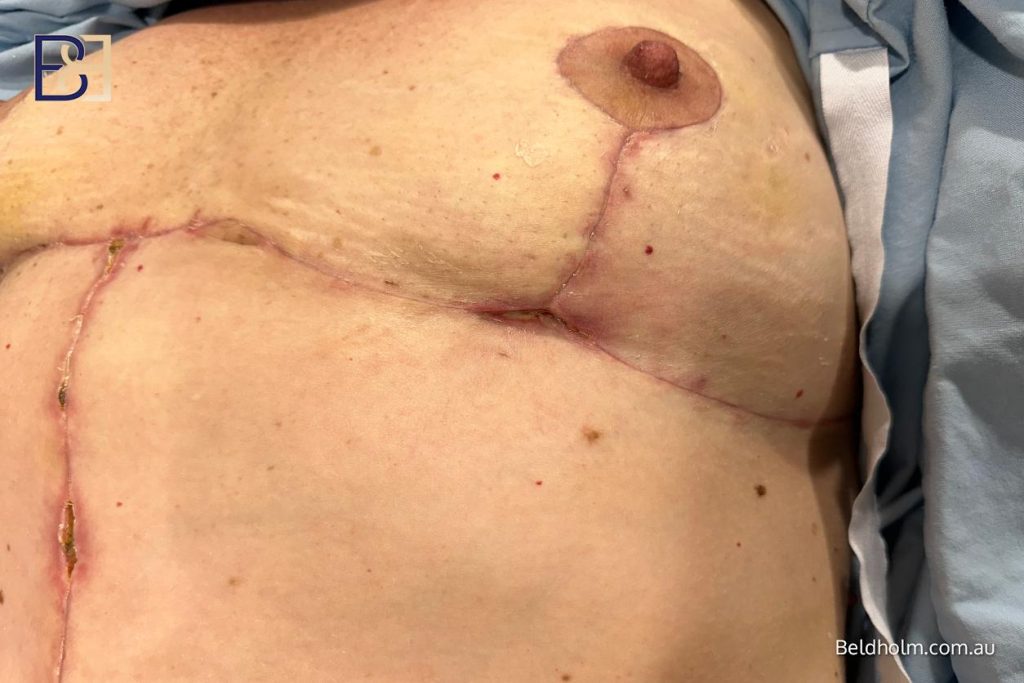
The incisions used in this operation are longer than those of a lift on its own, because the same cuts that reposition the nipple and reshape the breast also allow the flap of tissue to be moved. More incision means more wound to heal, and healing does not always run smoothly along its whole length.
The most common wound complication is a small area where the incision separates or is slow to close over. This is called dehiscence. It usually happens at the points where incision lines meet, because those junctions are under the most tension and have the most layers of tissue coming together. In the breast lift patterns used here, the two spots that most often give trouble are the junctions along the vertical scar and the point where the vertical and horizontal incisions meet at the base of the breast, sometimes called the triple point.
In the published auto augmentation series this is a recognised but minor issue:
- In a dermal-suspension series, minor separation at the triple point was seen in about five per cent of patients (3).
- In a mushroom-flap series, around nine per cent had delayed wound healing, most commonly at the upper and lower junctions of the vertical scar. All of these settled with dressings and healed without further surgery (1).
Most areas of delayed healing or minor separation are managed with dressing changes and time. They close on their own over days to weeks. A larger separation occasionally needs the wound cleaned and re-closed, but this is uncommon.
Two related points:
- Fluid collection. A seroma is a build-up of clear tissue fluid under the skin. Small ones settle on their own. A larger one may need to be drained with a needle in clinic.
- Wound support during healing. I use Comfeel dressings on the incisions. Keeping tension off the healing wounds in the early weeks helps the junctions hold.
Smoking has a strong effect on wound healing, because it reduces the blood supply that a healing wound and a repositioned flap both depend on. This is the main reason I ask patients to stop smoking for six weeks before and six weeks after surgery. Raised blood sugar in diabetes and poor nutrition also slow healing, which is why these are assessed and managed before surgery.
Nipple and areola complications

Moving the nipple and areola to a higher position is part of every breast lift. The nipple and areola stay attached to the breast through a bridge of tissue called a pedicle, and that pedicle carries the blood supply that keeps them alive and healthy. Protecting that blood supply is one of the central technical concerns of the operation.
Changes in nipple sensation
Sensation to the nipple and areola can change after surgery. It may be reduced, heightened, or patchy, and it can affect part or all of the nipple. In many patients sensation returns over the following months as the nerves recover, but some change can be permanent. Sensation is carried by nerves that run through the breast tissue, and repositioning the nipple can stretch or interrupt them. This is discussed with every patient before surgery, because it cannot be guaranteed either way.
Partial loss of the nipple or areola
The more serious concern is the blood supply itself. If the pedicle is compromised, part of the nipple or areola can suffer reduced blood flow. In its mildest form this shows as a change in the colour of the skin that recovers on its own. In more significant cases, a portion of the nipple or areola skin can be lost and needs time and dressings to heal, occasionally with a minor procedure to tidy the area.
Complete loss of the nipple is rare in this operation and was not seen in the published series discussed here. Partial complications are more relevant:
- In an inferior-pedicle auto augmentation series, one patient (around two per cent) had partial loss of the nipple-areola complex, which healed after a debridement and closure. The total rate of blood-supply-related complications, including one case of temporary pigmentation change in the areola, was under three per cent (2).
- In a mushroom-flap series, there were no cases of nipple-areola loss (1).
- In a dermal-suspension series, there were no cases of nipple loss (3).
How I manage this risk
I choose the pedicle to preserve a robust blood supply to the nipple, and I check the tissue for healthy bleeding during the operation before relying on it. During closure, if the nipple sits under tension, the tissue is released so it can sit without being pulled tight. If a blood-supply compromise does appear after surgery, early review gives the best chance of managing it well.
Fat necrosis and flap viability
This is the risk most specific to auto augmentation, because it relates directly to the tissue that is moved to add volume.
When I raise a flap of your own breast tissue and reposition it higher on the chest wall, that tissue has to keep its blood supply in its new position. It stays connected to the body through the pedicle, and as long as blood flows through it well, the tissue survives and integrates. The concern is what happens if part of that tissue does not get enough blood.
What fat necrosis is
Fat necrosis is the death of a small area of fatty tissue when its blood supply is not enough to keep it alive. It is not cancer and it is not dangerous in itself, but it can affect the result and cause concern until it is understood.
It usually shows up as a firm lump or a hardened area in the breast, sometimes weeks or months after surgery. The overlying skin can occasionally look different over the affected spot. A small area may soften and settle over time on its own. A larger or persistent area sometimes needs a minor procedure to remove it.
The reason it matters beyond the lump itself is that a firm area in the breast needs to be told apart from other causes. Imaging, usually an ultrasound and sometimes a mammogram, is used to characterise it. Occasionally a needle sample is taken to confirm what it is. This is a normal part of investigating any new firm area in the breast, and it is why any new lump after surgery should be checked rather than assumed to be scar tissue.
How likely it is
In the published auto augmentation series, significant fat necrosis was uncommon. In a dermal-suspension series, there were no cases of detectable fat necrosis, and flap survival was confirmed during the operation by checking for brisk bleeding at the tip of the moved tissue (3). Smaller areas of fat necrosis can occur with any operation that repositions breast tissue, and are more likely where blood supply is already under strain.
What raises the risk, and how I manage it

The chance of fat necrosis goes up when blood supply to the flap is reduced. The main avoidable factor is smoking, which narrows blood vessels and cuts the blood flow the flap depends on. This is a further reason smoking cessation matters before and after surgery. Larger flaps and more extensive repositioning also place more demand on the blood supply.
Scarring
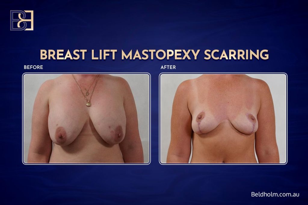
This operation leaves permanent scars, and they are more extensive than the scars of a lift on its own. The incisions do two jobs at once: they reposition the nipple and remove excess skin, and they allow the flap of tissue to be raised and moved to add upper pole volume. That extra work means more incision.
Where the scars sit
Disclaimer: Results vary, surgery has risks, seek second opinion. Operation performed by Dr Beldholm.
The pattern depends on how much lift and reshaping is needed. In most cases the scars follow a lift pattern:
- Around the edge of the areola
- A vertical line running down from the areola to the crease under the breast
- In many cases, a horizontal line along the crease under the breast, giving an anchor or inverted-T shape
Some patients have a shorter pattern without the horizontal component, sometimes called a lollipop scar. Which pattern applies is determined by your anatomy and what the operation needs to achieve, and it is discussed before surgery so you know what to expect.
How scars mature
Fresh scars are firm, raised and pink or red. Over roughly twelve to eighteen months they usually flatten, soften and fade towards a paler line. They never disappear completely. Final scar appearance varies a great deal between people and is influenced by your skin type, your genetics and how your body heals, none of which can be fully predicted in advance.
Thickened and raised scars
Some people form scars that stay thick, raised or firm rather than settling. A hypertrophic scar is a raised scar that stays within the line of the incision. A keloid scar extends beyond it. These are more common in some skin types and family backgrounds.
In the published auto augmentation series, troublesome scarring was uncommon. In a mushroom-flap series, one patient developed a hypertrophic scar that was treated with a corticosteroid injection (1). Where a scar stays thick, options include corticosteroid treatment, silicone therapy, and other scar management measures.
Scar revision
A proportion of patients choose to have a scar revision with a further minor procedure once it has matured. In the same mushroom-flap series, around nine per cent of patients had an aesthetic reintervention for scar correction, done under local anaesthetic (1). A revision can improve a scar but cannot erase it.
The trade-off
The scar burden is the main trade-off of this operation. It buys a lift and added upper pole volume from your own tissue, but it costs a longer scar than a lift alone. In the published experience, patients who understood this in advance and accepted the scars. Being clear about the scars before you decide is part of making that a decision you are comfortable with.
I place incisions along the standard lift patterns so the scars fall where they are most easily concealed, close carefully to keep tension off the healing wounds, and review your scars through the follow-up period so any thickening is picked up and managed early.
Asymmetry, shape change and recurrent ptosis
Breasts are rarely identical to begin with, and no operation makes the two sides an exact match. Some breast asymmetry in size, shape or nipple position between the two sides is normal before surgery and can remain after it. The aim is to improve the balance.
Asymmetry after surgery
Even when the same operation is done on both sides, the two breasts can heal differently. One may settle slightly higher, fuller or firmer than the other. Small differences are common and usually do not need anything done. A more noticeable difference sometimes leads a patient to choose a further procedure to improve the match. To reduce this, I perform the shaping and suspension on both sides together during the operation, and check symmetry with the patient sitting upright rather than lying flat, because the breast sits differently in each position.
How the shape settles over time
The shape you see immediately after surgery is not the final shape. Over the first several months the tissue settles, swelling resolves, and the breast takes on its longer-term form. The upper pole fullness that this operation adds can change as the tissue settles and as gravity acts on it over the following months and years. This is a normal part of how the breast behaves, not a failure of the operation, but it means the early result and the settled result are not the same thing.
Recurrent ptosis
Ptosis is the medical term for a lower breast position, where the breast tissue and nipple sit lower on the chest. A lift raises the tissue and nipple to a higher position. Over time, some downward settling can return. This is called recurrent ptosis, and how much occurs varies with your skin elasticity, breast size and volume, and the demands that gravity and time place on the tissue.
The published auto augmentation series report on durability in different ways:
- A dermal-suspension series suspends the breast tissue to the covering of the rib to hold the lift, and reports that recurrence of a lower position is less likely with this anchoring than with tissue-only techniques (3).
- A mushroom-flap series reported that the secondary settling seen in many patients around six months after a traditional lift was not seen in their series over the follow-up period, though long-term data beyond two years is not yet available (1).
No lift lasts forever, and none of these techniques stops the ageing of breast tissue. What a durable technique aims to do is hold the position for as long as possible.
Further surgery
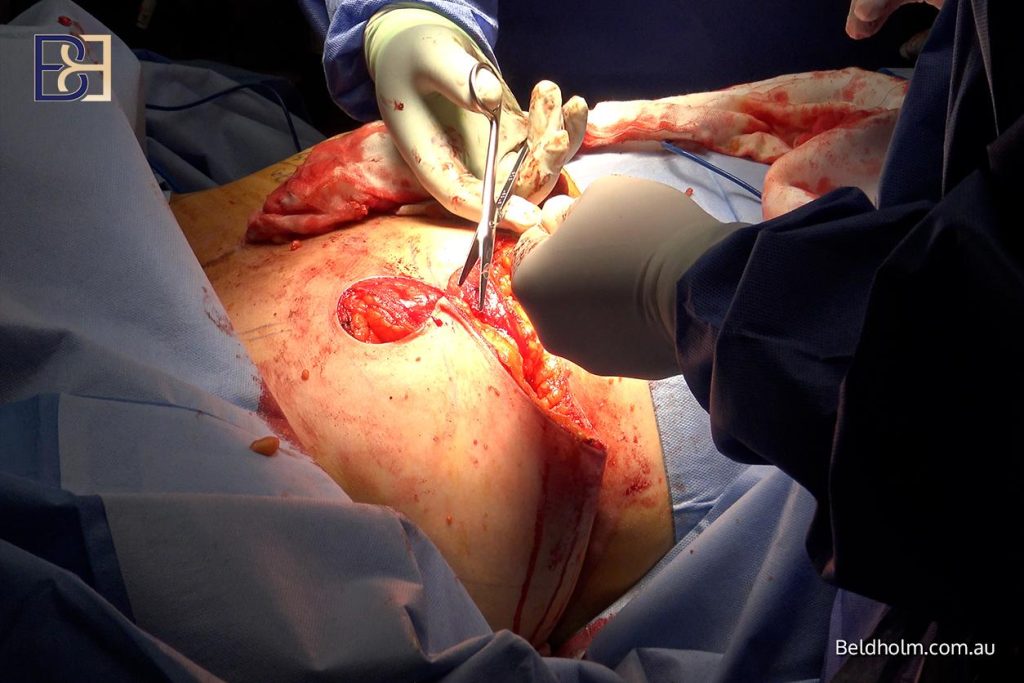
A proportion of patients choose a further procedure, either to treat symmetry or to refine the result. This kind of revision surgery is uncommon but does happen. In a mushroom-flap series, around nine per cent of patients had a further aesthetic procedure, most commonly for scar correction (1). In an inferior-pedicle series, around six per cent had a second lift because the first had not lifted as much as intended (2).
The prospect of possible further surgery is part of the picture to weigh before the first operation. Choosing a technique with good durability, and being realistic about what a single operation can achieve, both reduce the chance of needing more.
Effect on future breastfeeding, and the effect of future pregnancy
This operation moves breast tissue and repositions the nipple and areola, so it is reasonable to ask what it means for breastfeeding and for how the result holds if you have a child afterwards. Not every patient is planning a pregnancy, and this section applies only if that is relevant to you.
Breastfeeding after this operation

Breastfeeding depends on the milk-producing tissue and its ducts staying connected to the nipple, and on the nerve supply to the nipple that triggers milk release. Any operation that repositions the nipple and reshapes the breast has the potential to affect this, because the ducts and nerves run through the tissue that is moved.
Many people can still breastfeed after breast surgery, but it cannot be guaranteed, and some find their supply is reduced. How much effect there is depends on the technique used and on your own anatomy, and it cannot be predicted precisely in advance. If breastfeeding a future child matters to you, tell me at consultation, because it is part of the assessment and the discussion about whether and how to proceed.
The effect of a future pregnancy on the result

Pregnancy and breastfeeding change the breast. The tissue enlarges and then reduces again afterwards, and breast volume, shape and position can be different once things settle. If you have this operation and then become pregnant, those changes can alter the result you were left with, including the lift and the added upper pole volume.
For that reason, where a future pregnancy is part of your plans, surgery is generally planned once your family is complete and breastfeeding has finished, so the result is not undone by later changes. This is a timing judgement made individually at consultation, not a fixed rule or a set interval. Some patients proceed earlier for their own reasons, understanding that a later pregnancy may change the outcome.
The point of raising this is not to delay anyone unnecessarily. It is so the decision about when to operate is made with a clear picture of how a later pregnancy can affect what the operation achieves.
Factors that increase your risk
Some of the risks on this page are more likely in certain circumstances. Several of those circumstances can be changed before surgery, and part of preparing for the operation is working through them. None of this is about ruling people out. It is about knowing what raises the risk so it can be managed.
Smoking
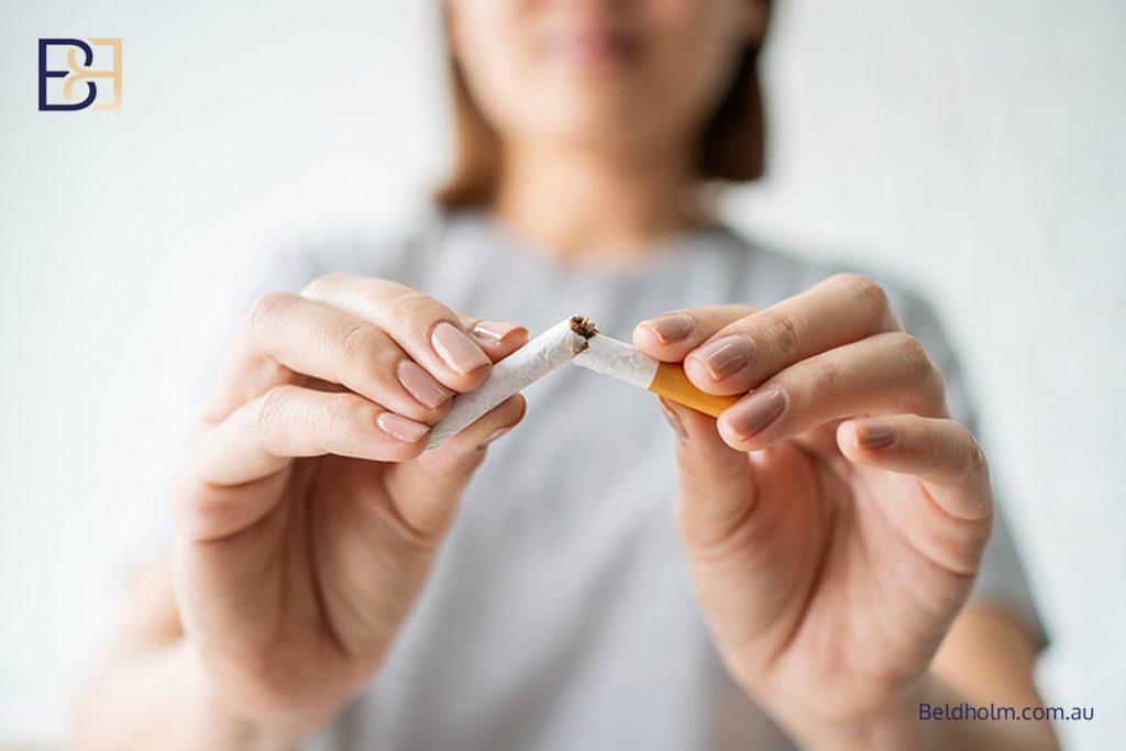
Smoking is the single most important avoidable risk factor for this operation. It narrows blood vessels and reduces the blood flow that healing wounds, the repositioned flap, and the nipple and areola all depend on. That raises the risk of wound healing complications, fat necrosis, and reduced blood supply to the nipple. I ask patients to stop smoking for six weeks before and six weeks after surgery. This includes vaping and nicotine replacement, because nicotine itself is what reduces the blood flow.
Raised body weight

A higher body weight is associated with more wound healing and anaesthetic risk across all surgery. In the published auto augmentation experience, some series set an upper weight or body mass index limit for the operation. In my practice, body weight is one part of the overall assessment rather than a single cut-off number, and it is considered alongside your general health and the specifics of your case.
Diabetes and blood sugar control
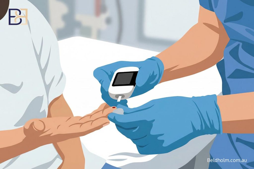
Diabetes, particularly when blood sugar is not well controlled, slows wound healing and raises infection risk. Where diabetes is present, getting blood sugar into good control before surgery is part of the preparation, and it is assessed as part of deciding whether and when to proceed.
Previous breast surgery or scars
Previous surgery on the breast can alter the tissue and its blood supply, which affects how a flap can be raised and how the tissue heals. Existing breast scars are noted at assessment because they change the surgical picture.
Active skin conditions
Active skin conditions in the area, such as significant inflammation in the fold under the breast, raise the risk of wound healing trouble and infection. These are usually settled before surgery rather than operated through.
General health and anaesthetic fitness
Your overall fitness for an anaesthetic matters, because this is an operation under general anaesthetic. Significant heart, lung or other medical conditions are assessed before surgery, and some conditions need to be stable and well managed before an operation of this size is appropriate.
None of these factors is a straightforward yes or no. They are weighed together at consultation, alongside your medical history and after a GP referral, to work out whether the operation is appropriate for you and how to make it as low-risk as it reasonably can be.
Warning signs and when to contact us

Most recovery runs without serious trouble, but knowing what to watch for means a complication is picked up and dealt with early. Contact us if you notice any of the following.
- One breast becoming rapidly more swollen, firmer, more painful or more bruised than the other, particularly in the first day or two. This can signal a haematoma.
- Spreading redness, increasing warmth, worsening pain rather than settling pain, or fluid or pus draining from a wound. These can signal infection.
- A fever.
- A part of the nipple or areola turning dark, dusky or pale and not recovering. This can signal a blood supply concern and should be checked without delay.
- A wound edge separating, or an area that will not close over.
- A new firm lump in the breast. Most are not serious, but any new lump after surgery should be checked rather than assumed to be scar tissue.
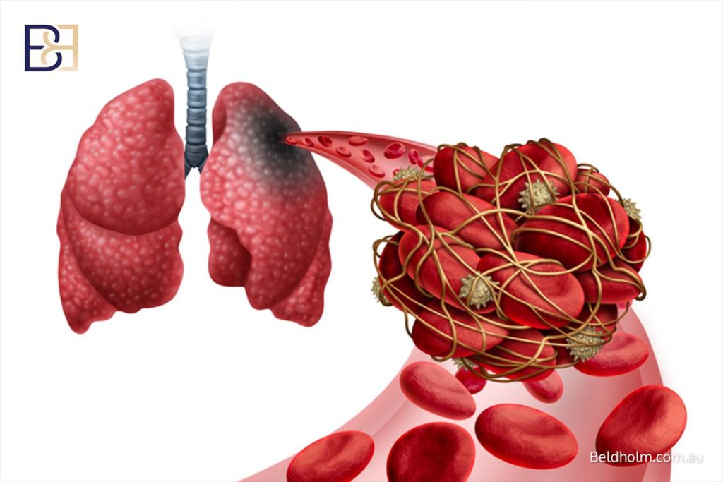
Some symptoms need urgent attention on their own. Chest pain, difficulty breathing, or a hot, swollen, painful calf can point to a blood clot, including a deep vein thrombosis or a pulmonary embolism, and are treated as an emergency.
How to reach the right care
- During clinic hours, call the rooms. The team will advise and arrange review as needed.
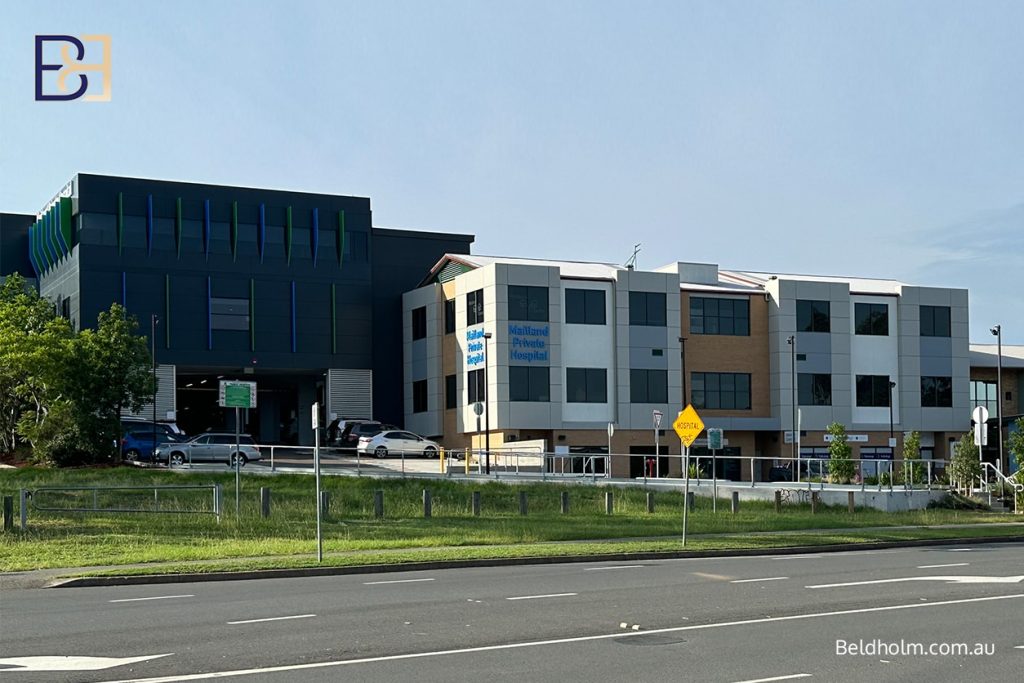
- After hours, call Maitland Private Hospital. The nursing team provides phone triage and will guide you on what to do next.
- If a wound or physical symptom needs to be examined in person and it cannot wait, go to your local emergency department. Maitland Private is not an emergency department.
- For anything life-threatening, including chest pain or difficulty breathing, call 000.
When in doubt, make contact. It is always better to have something checked and found to be minor than to leave a genuine complication unattended.
A final word on the risks

Everything on this page is here so you understand what can happen after this operation and know what to act on. For most people the complications described here are either uncommon or manageable when they are picked up early, which is why the warning signs above matter and why the follow-up reviews are built into your care.
Results vary from patient to patient, and nothing here predicts your individual outcome. If you have had this operation and something does not seem right, use the contacts above rather than waiting. If you are preparing for surgery and want to go through any of these risks in more detail as they apply to you, we can do that at your appointments.
References
- Van Boeckel V, Nizet C, Nizet JL, Nelissen X. Retrospective multicenter review of mastopexy using the “mushroom” auto-augmentation flap technique. Plast Reconstr Surg Glob Open. 2026;14(5):e7774.
- Kirwan L, Wazir U, Mokbel K. Breast auto-augmentation: a versatile method of breast rehabilitation, a retrospective series of 107 procedures. Arch Plast Surg. 2015;42(4):438-445.
- Rubin JP, Khachi G. Mastopexy after massive weight loss: dermal suspension and selective auto-augmentation. Clin Plast Surg. 2008;35(1):123-129.