Gynaecomastia means enlarged male breast tissue. The surgery to remove excess tissue is sometimes called male breast reduction, or male breast reduction surgery. When surgeons talk about it, they usually describe it in grades, from mild through to severe. The grade is a shorthand for how much excess breast tissue is present, whether there is excess skin, and where the nipple sits. It is a medical condition, not a cosmetic label, and it is graded so the surgical procedure can be matched to what is actually there.
The trouble is that there is no single agreed grading system. Several have been published over the last sixty years, and they do not all use the same definitions. Different websites quote different grades, different treatments, and different cut-off points, which is confusing if you are trying to understand your own situation.
I have gone back to the original research papers for each of the major systems and set them out here side by side. For each one I have included the grades as the authors defined them, along with the treatment each author recommended at the time. Several of these systems are old, and the treatments described in them are not what I would do today. I have flagged this clearly. These are the original authors’ recommendations, not mine.
My aim here is to clear up the confusion, show how thinking about gynaecomastia has changed over time, and explain how grade influences the operation. Whether surgery is appropriate for any individual, and which technique suits them, is decided at consultation after examination and the appropriate workup. A GP referral is needed before any consultation, and results vary from patient to patient.
Gynaecomastia and pseudogynaecomastia: gland versus fat
Before any grading system makes sense, it helps to understand what is actually being graded. The male chest can enlarge for two different reasons, and telling them apart matters because it changes the operation.
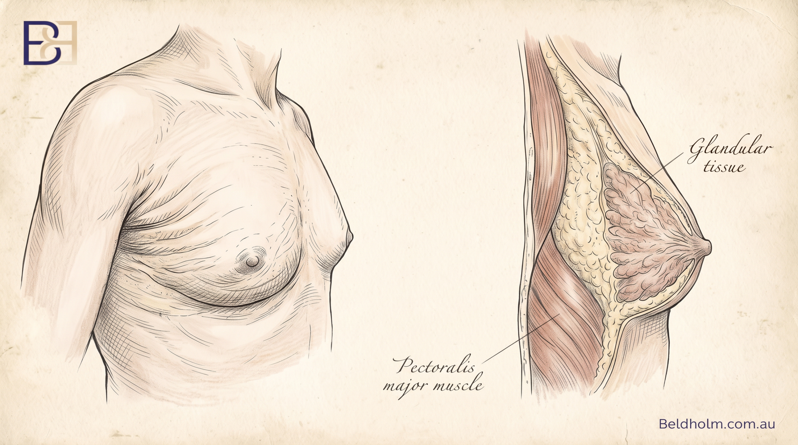

Gynaecomastia (true gynaecomastia) is enlargement of the glandular breast tissue. It is driven by a shift in the balance between oestrogen and testosterone, so that oestrogen activity is relatively higher. This can happen at several points in life, and there is more than one possible cause. Common ones include the normal hormonal changes of puberty, the hormonal changes of older age, certain medications, anabolic androgenic steroids, and some underlying medical conditions. In a proportion of men no cause is ever found, which is called idiopathic gynaecomastia (1). The firm glandular disc sits directly under and around the nipple, and you can usually feel it as a distinct rubbery layer.
Pseudogynaecomastia (lipomastia) is enlargement caused by excess fat rather than gland. The chest looks fuller, but there is no firm disc under the nipple. This is common with weight gain and is being seen more often after massive weight loss, where excess skin adds to the appearance. Here the enlarged breast tissue is fat tissue, not glandular tissue.
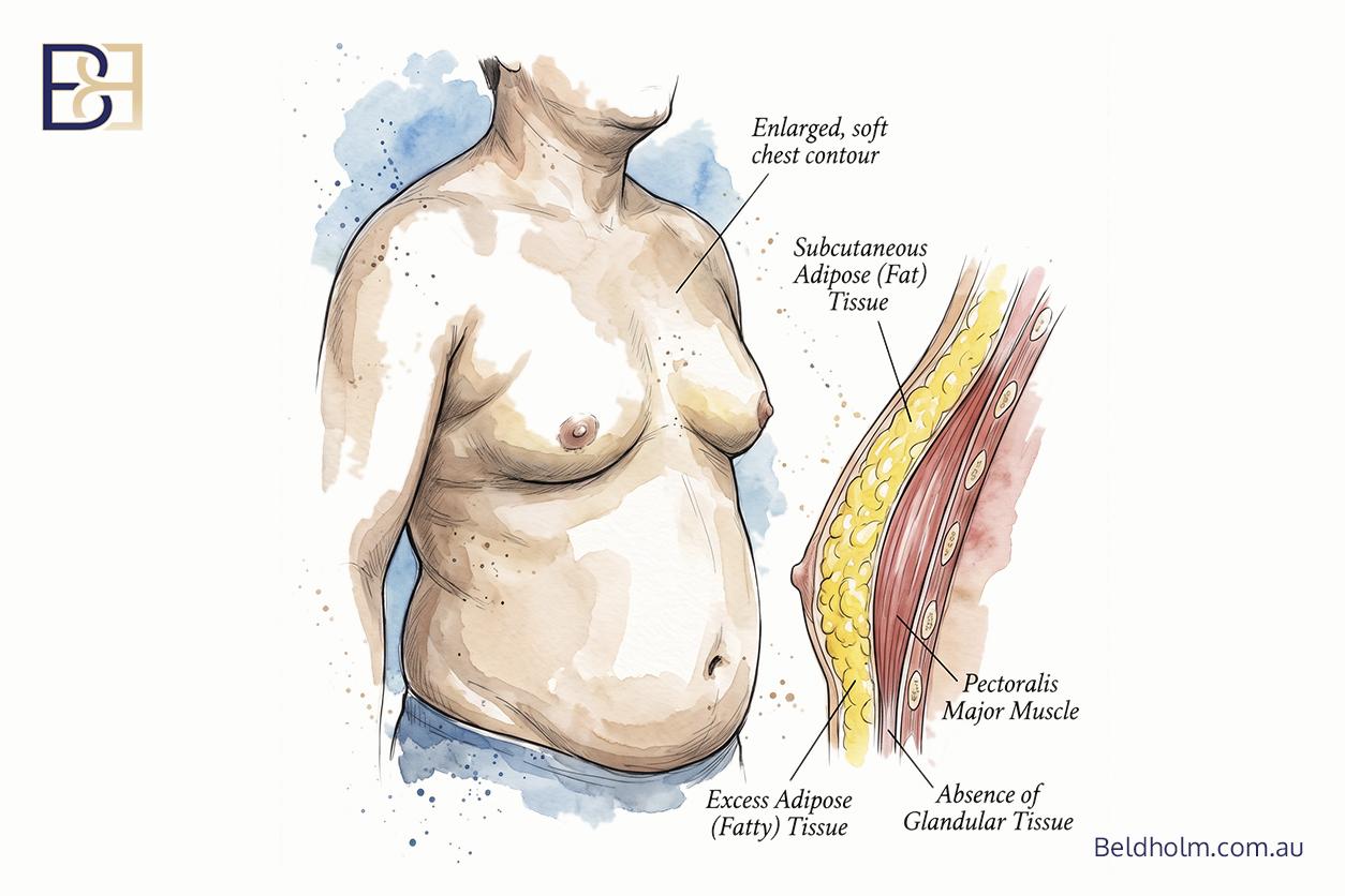
Many men have a mix of both, some firm glandular breast tissue and some excess fatty tissue. Enlarged male breasts are rarely one thing or the other.
Why the distinction drives technique
This is the single most important reason grade alone does not tell the whole story. Liposuction removes fat. It does not reliably remove the firm glandular disc. So a chest that is mostly fat may respond to liposuction, while a chest with a genuine glandular component usually needs that disc removed directly through a small incision, a procedure called glandular excision or subcutaneous mastectomy. Getting this wrong is a common reason men are unhappy after surgery, because the fat was suctioned but the firm lump under the nipple was left behind.
Where anabolic androgenic steroids are the driver, the point is clinical rather than anything else. Steroid-related gynaecomastia tends to be more glandular, and it can settle once the agent is stopped under medical guidance (2). If the agent is still being used, that is managed first, because operating around an active cause raises the chance the tissue comes back. I do not give any advice on using or obtaining these agents. What matters here is that an ongoing driver affects both suitability and the risk of recurrence.
A proper examination sorts out how much of each is present. That assessment, not a photo or a grade number, is what determines the operation.
Table of Contents
Why grading systems exist, and what they can and cannot do
Grading systems were created to give surgeons a shared language. If one surgeon says a patient has Grade III gynaecomastia, another surgeon should have a rough idea of what that means without seeing the patient. The grade is also meant to help match the operation to the severity, so that milder cases are not over-treated and more advanced cases are planned properly.
Used that way, grading is useful. It supports clear communication, it helps with surgical planning, and it helps set realistic expectations about the size of the operation and the scars.
What grading does not do
There are two things a grade cannot tell you.
First, a grade does not predict whether gynaecomastia will settle on its own. That depends far more on the cause and how long it has been present than on how large it is. Recent pubertal change often settles without any surgery. Steroid-related change may settle once the agent is stopped. A grade is a snapshot of size and shape, not a prediction.
Second, a grade does not replace working out the cause. An underlying or reversible cause is investigated and treated before surgery is considered, because surgery does not change an ongoing driver. Where a hormonal cause, a medication, or an agent is still active, the changes can return even after a technically good operation. This is why the workup comes first and the grade comes second.
Working out the cause

Because the cause matters more than the grade, the workup comes before any talk of a surgical procedure. What that involves depends on the history and examination, but it commonly includes:
- Medical history. Medications, anabolic androgenic steroids, alcohol, recreational drugs, and any change in weight. Some medications are a recognised cause, and where one is implicated, it is reviewed with the doctor who prescribed it. Nobody should stop a prescribed medication on their own.
- Blood tests. Hormone levels, including testosterone, oestradiol, LH, FSH, prolactin and beta-hCG, are checked where indicated to look for a hormonal cause. Liver and kidney function and thyroid function are also relevant, because liver disease, kidney disease and thyroid disorders can all drive enlarged breast tissue.
- Examination for anything that does not fit. This matters. Male breast cancer is rare, but it exists, and it does not present like gynaecomastia. A firm or hard lump that sits off to one side rather than centrally behind the nipple, a lump in one breast only, skin dimpling, nipple retraction, bleeding from the nipple, or an enlarged lymph node in the armpit are all findings that need investigating rather than grading. Where there is any doubt, imaging and specialist referral come first. No grading system substitutes for excluding breast cancer.
Some causes can be treated without an operation at all. Where a drug is the driver and can be changed, or where a hormonal or medical condition is treatable, non surgical treatments are the appropriate first step, and in some men the enlargement settles once the cause is treated.
So a grading system is a planning tool, not a decision-maker. It helps describe what is there and guide technique. It does not decide, on its own, whether someone should have surgery.
How grade is assessed in practice
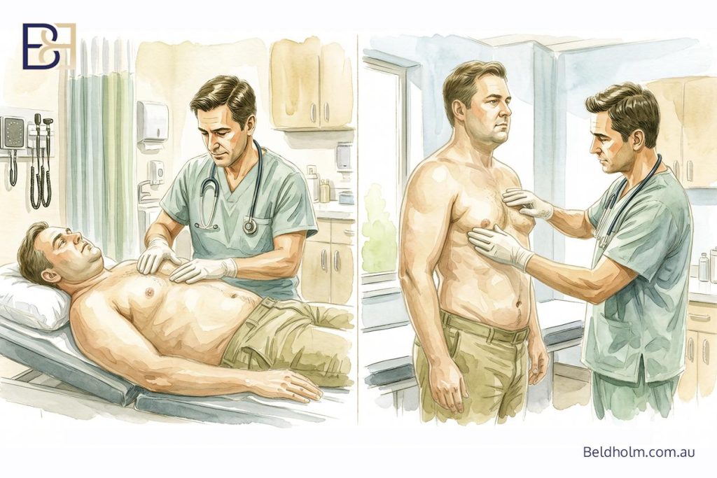
Grading is done at a physical examination, not from a photograph. There are a few things I am assessing when I examine a chest.
- The firm disc. I palpate under and around the nipple to feel whether there is a firm glandular disc, and how large it is. This is what separates true gynaecomastia from a chest that is mostly fat.
- The amount of tissue. How much excess breast tissue is present overall, glandular tissue and fat tissue together, and whether one side is larger than the other.
- Skin excess. Whether the skin has been stretched and how well it is likely to redrape once I remove excess tissue. Good skin elasticity usually means the skin will retract on its own. Poor elasticity means excess skin has to be removed. Skin quality matters more in men who have had massive weight loss.
- Nipple position. Where the nipple and areola sit, and whether they have descended. Different published systems use different landmarks for this, which I will come back to when I compare them.
The reason this has to be done in person is that the firmness of the tissue and the quality of the skin cannot be judged from an image. Two chests that look identical in a photo can need different operations once you feel the tissue.
This is also why a grade is a starting point for a conversation, not a decision made before you are seen. Suitability and technique are worked out in person, after examination and any workup needed to identify a cause.
The published classification systems, in order
Below are the major published gynaecomastia classification systems, set out from the earliest to the most recent. This is the evolution of the idea, from a basic measure of gland size in 1961 through to systems that account for skin quality and post-weight-loss presentations.
For each system I have listed the grades as the authors defined them, and the treatment each author recommended. A few points to keep in mind as you read them:
- These are the original authors’ recommendations, not mine. Several of these systems are decades old and the treatments in them are not what I would do today. I have kept them as published so you can see how thinking has changed.
- Each system uses its own landmarks. Where a system measures nipple position against the inframammary fold (the crease under the chest), that is the landmark those authors chose. It is not necessarily the one I use, and I explain my own approach separately.
- Grade does not decide the operation on its own. As above, the cause and the examination matter as much as the grade.
Each system also has its own detailed article, linked at the end of its section.
Nydick classification (1961)
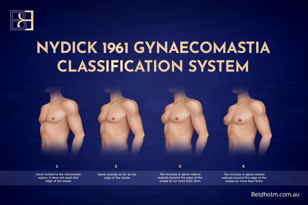
The Nydick system was one of the earliest attempts to grade male breast enlargement (3). It came out of a study of adolescent boys and it is built entirely around the diameter of the glandular tissue, measured against the edge of the areola. It does not account for skin excess or nipple position, which makes it far simpler than anything that came later.
Its main value now is historical, and its central message still holds: the study followed pubertal gynaecomastia and found it commonly settled on its own, so the authors recommended observation rather than surgery in adolescents.
Nydick grades
- Grade 1: Gland limited to the area behind the areola, not reaching the edge of the areola.
- Grade 2: Gland extends as far as the edge of the areola.
- Grade 3: Gland extends beyond the edge of the areola by no more than 5 mm.
- Grade 4: Gland extends beyond the edge of the areola by more than 5 mm.
Treatment the authors recommended: observation only, across all grades, on the basis that adolescent gynaecomastia usually settles without treatment.
Read the full breakdown in the dedicated article: Nydick 1961 classification article.
Simon classification (1973)
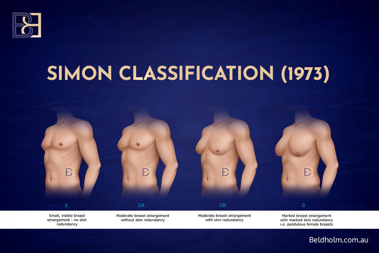
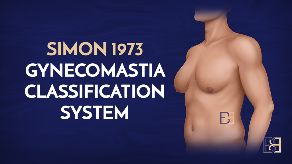
The Simon system is the one most surgeons and most websites still refer to (4). It was an important step forward because it graded the condition from a surgical point of view, taking both breast size and skin excess into account. It also introduced the idea of subdividing the moderate grade based on whether skin excess is present, which is a distinction that still drives technique today.
Simon grades
- Grade 1: Small, visible enlargement with no skin excess.
- Grade 2A: Moderate enlargement with no skin excess.
- Grade 2B: Moderate enlargement with skin excess.
- Grade 3: Marked enlargement with significant skin excess and ptosis.
Treatment the authors recommended
- Grade 1 and 2A: Subcutaneous mastectomy through an infra-areolar incision.
- Grade 2B: Subcutaneous mastectomy through a supra-areolar incision, with excision of skin above the areola in a semicircle.
- Grade 3: Excision of skin as well as gland, with a staged approach suggested.
The strength of the Simon system is its simplicity, which is also its limitation. It does not separate glandular from fatty tissue, and it predates modern liposuction, so it does not tell you when liposuction alone might be enough.
Read the full breakdown in the dedicated article: Simon 1973 classification article.
Cohen classification (1987)
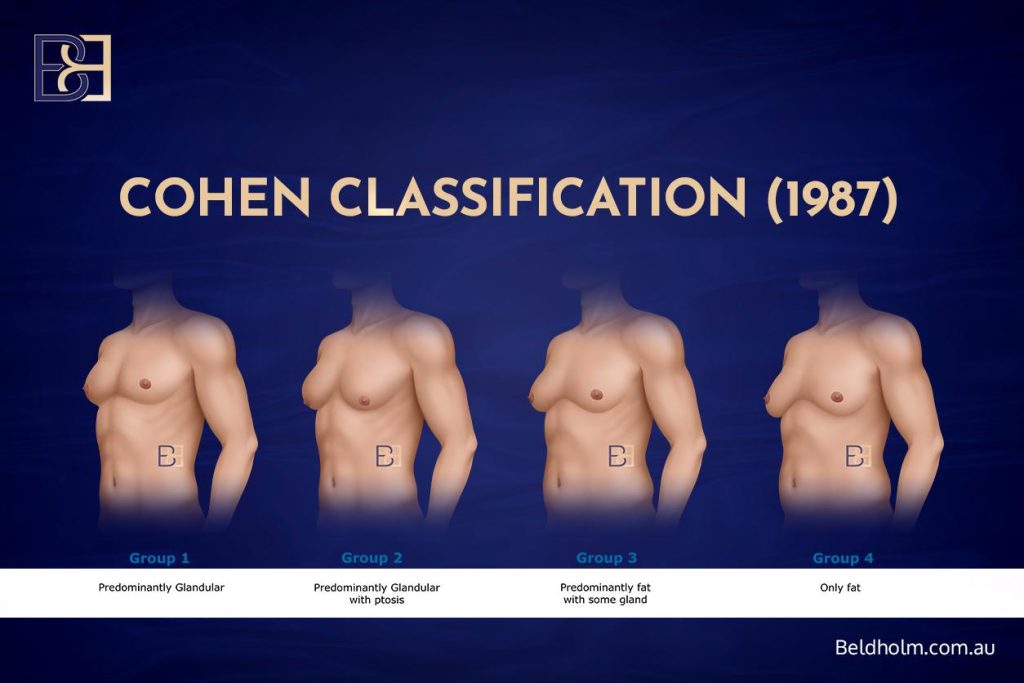
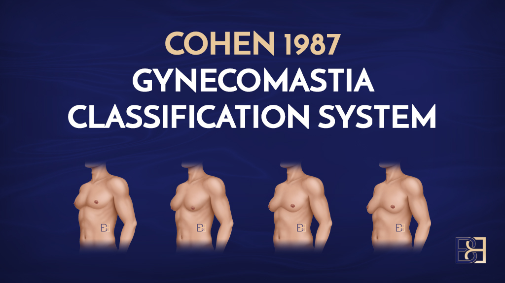
The Cohen system is one of the more straightforward ones (5). Rather than grading by size, it groups patients by what the tissue is made of, gland versus fat, and whether ptosis is present. That makes it a practical way to decide whether someone needs skin excision or can be managed with less invasive techniques.
Cohen groups
- Group 1: Predominantly glandular.
- Group 2: Predominantly glandular with ptosis.
- Group 3: Predominantly fat with some gland.
- Group 4: Fat only.
Treatment the authors recommended
- Group 1: Open excision.
- Group 2: Open excision and liposuction, with sharp dissection to release the inframammary fold.
- Group 3: Open excision and liposuction.
- Group 4: Liposuction only.
The appeal of the Cohen approach is that it puts the gland-versus-fat question front and centre, which is the distinction that matters most for technique. What it does not do is grade severity in much detail.
Read the full breakdown in the dedicated article: Cohen 1987 classification article.
Rohrich classification (2003)
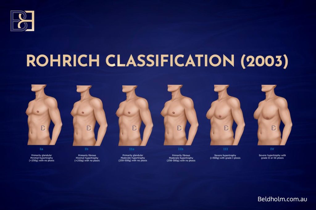
The Rohrich system was designed around the role of liposuction, and specifically ultrasound-assisted liposuction (6). It grades by the degree of enlargement measured by tissue weight, and it adds a note of whether the tissue is primarily glandular or primarily fibrous, then factors in ptosis at the higher grades. It is a useful system for planning when liposuction is central to the operation.
Rohrich grades
- Grade Ia: Minimal enlargement (under 250 g), primarily glandular, no ptosis.
- Grade Ib: Minimal enlargement (under 250 g), primarily fibrous, no ptosis.
- Grade IIa: Moderate enlargement (250 to 500 g), primarily glandular, no ptosis.
- Grade IIb: Moderate enlargement (250 to 500 g), primarily fibrous, no ptosis.
- Grade III: Severe enlargement (over 500 g) with grade I ptosis.
- Grade IV: Severe enlargement with grade II or III ptosis.
Treatment the authors recommended
- Grades I and II: Ultrasound-assisted liposuction.
- Grades III and IV: Ultrasound-assisted liposuction, with or without staged excision, the staged excision performed six to nine months after the liposuction.
The Rohrich system reflects the era when ultrasound-assisted liposuction was being defined as a tool for gynaecomastia. Its weakness for higher grades is that relying on liposuction with delayed excision does not suit every patient, particularly where a firm glandular disc or significant skin excess is present.
Read the full breakdown in the dedicated article: Rohrich 2003 classification article.
Cordova and Moschella classification (2008)
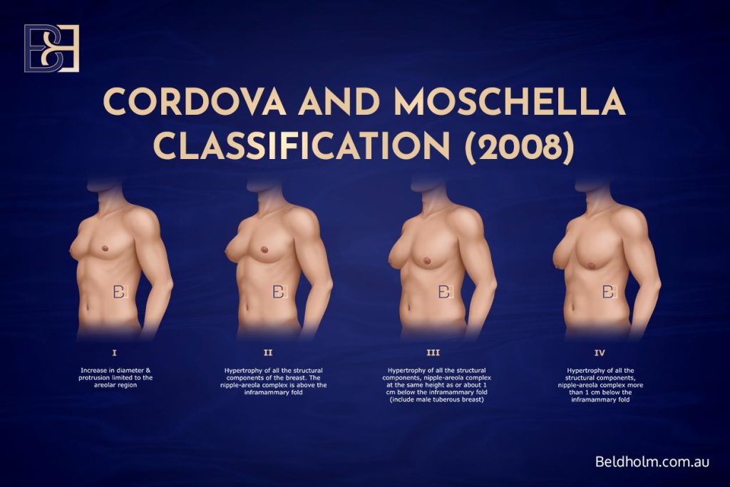
The Cordova and Moschella system grades by the combination of tissue type, glandular and fatty, and skin quality, using nipple position against the inframammary fold as the marker of severity (7). It is a structured, surgically focused system.
Cordova and Moschella grades
- Grade I: Increase in diameter and protrusion limited to the areolar region.
- Grade II: Enlargement of all the structural components of the breast, with the nipple-areola complex above the inframammary fold.
- Grade III: Enlargement of all components, with the nipple-areola complex at the same height as, or about 1 cm below, the inframammary fold. This grade includes the male tuberous breast.
- Grade IV: Enlargement of all components, with the nipple-areola complex more than 1 cm below the inframammary fold.
Treatment the authors recommended
- Grade I: Subcutaneous mastectomy through an infra-areolar incision. Liposuction not required.
- Grade II: Ultrasound-assisted liposuction first, followed by subcutaneous mastectomy through an infra-areolar incision.
- Grade III: Ultrasound-assisted liposuction, then subcutaneous mastectomy, then circular removal of skin around the nipple-areola complex with a round-block suture.
- Grade IV: Central pedicle reduction techniques, without liposuction.
The Cordova and Moschella system is a good example of matching an escalating operation to escalating severity, and it makes the point that the highest grades usually need skin removal rather than liposuction.
Read the full breakdown in the dedicated article: Cordova and Moschella 2008 classification article.
Gusenoff classification (2008)
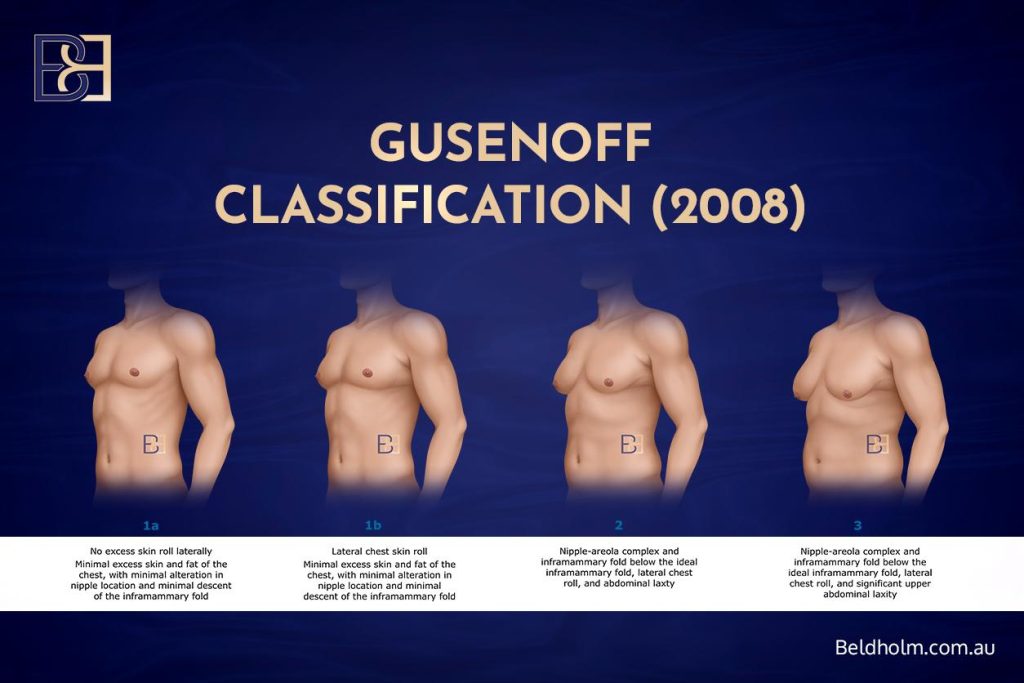
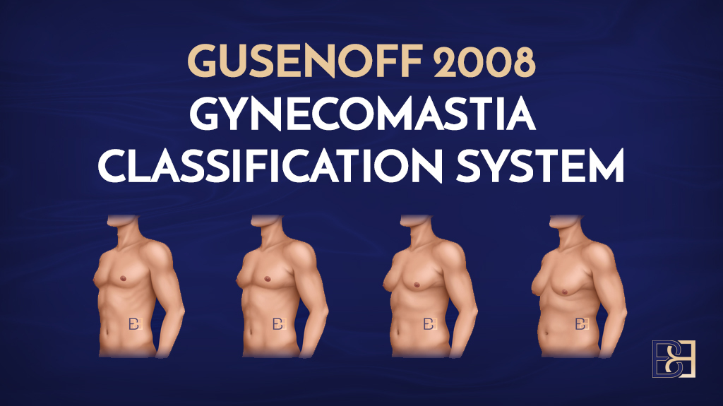
The Gusenoff system is different from the others because it was built specifically for men who have lost a large amount of weight (8). In this group the problem is often not a firm glandular disc but excess skin and fat, a lateral chest roll, and a nipple and inframammary fold that have dropped below where they should sit. The system grades those features.
Gusenoff grades
- Grade 1a: Minimal excess skin and fat, minimal change in nipple position and minimal descent of the inframammary fold, with no excess skin roll to the side.
- Grade 1b: As above, but with a lateral chest skin roll.
- Grade 2: Nipple-areola complex and inframammary fold sitting below the ideal position, with a lateral chest roll and minimal upper abdominal laxity.
- Grade 3: As grade 2, but with significant upper abdominal laxity.
Treatment the authors recommended
- Grade 1a: Liposuction (ultrasonic liposuction followed by suction-assisted lipectomy).
- Grade 1b: Liposuction with direct excision of the roll.
- Grade 2: Pedicled nipple reconstruction.
- Grade 3: Free nipple graft reconstruction.
The Gusenoff system is the one that best reflects the reality of post-weight-loss chests, where skin is the main issue. It is also the source I draw on for a more reliable landmark than the inframammary fold, which I discuss in my own approach below.
Read the full breakdown in the dedicated article: Gusenoff 2008 classification article.
Ratnam classification (2009)
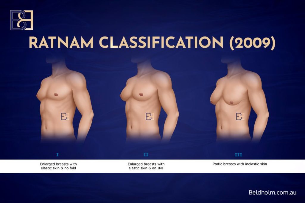
The Ratnam system grades by the amount of breast tissue, the degree of skin excess, and ptosis, and it is particularly focused on skin elasticity (9). It is a practical system for surgical planning because it makes clear when skin excision becomes necessary as the grades climb.
Ratnam grades
- Grade I: Enlarged breasts with elastic skin and no inframammary fold.
- Grade II: Enlarged breasts with elastic skin and a defined inframammary fold.
- Grade III: Ptotic breasts with inelastic skin.
Treatment the authors recommended
- Grade I: Subcutaneous mastectomy through an infra-areolar incision, with ultrasound-assisted liposuction.
- Grade II: As grade I, plus release of the inframammary fold with a blunt liposuction cannula, wide-area ultrasound-assisted liposuction, and treatment of the underside of the skin with ultrasonic energy to encourage skin tightening.
- Grade III: As grade II, plus reduction of the excess skin by de-epithelialisation around the areola and a purse-string closure.
The Ratnam system is useful for its attention to skin quality, which is a genuine determinant of outcome. Some of its specific techniques are less commonly used now, but the principle that inelastic, ptotic skin needs to be managed directly is sound.
Read the full breakdown in the dedicated article: Ratnam 2009 classification article.
Barros classification (2012)
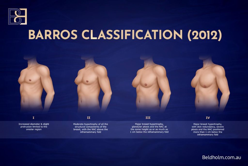
The Barros system grades by the shape, size, and degree of enlargement along with skin laxity, using nipple position against the inframammary fold as the marker of severity (1). It was designed to help with surgical planning and to guide whether limited tissue removal or a more extensive operation is needed, and it pays particular attention to skin laxity, which makes it relevant to weight-loss patients.
Barros grades
- Grade I: Increased diameter and slight protrusion limited to the areolar region.
- Grade II: Moderate enlargement of all the structural components of the breast, with the nipple-areola complex above the inframammary fold.
- Grade III: Major enlargement with glandular ptosis, and the nipple-areola complex at the same height as, or up to 1 cm below, the inframammary fold.
- Grade IV: Major enlargement with skin redundancy, severe ptosis, and the nipple-areola complex more than 1 cm below the inframammary fold.
Treatment the authors recommended
- Grade I: Subcutaneous mastectomy through an infra-areolar incision. Liposuction not required.
- Grade II: Liposuction first, then removal of the gland through an infra-areolar incision.
- Grade III: Liposuction first, then gland removal, with a donut mastopexy as part of the operation.
- Grade IV: Reduction with a superior or superomedial pedicle.
The Barros system is a well-rounded one that carries the escalation of technique through to skin excision at the top grades. It shares the limitation of most fold-based systems, which is that the inframammary fold is not always a reliable landmark, something I return to below.
Read the full breakdown in the dedicated article: Barros 2012 classification article.
Punia and Gupta classification (2023)

The Punia and Gupta system is the most recent of the major published systems (10). It builds on the Simon classification and puts more emphasis on skin redundancy and the degree of enlargement, and it recognises skin excess at an earlier grade, which helps decide when skin tightening is needed alongside tissue removal. It also brings liposuction, including around the axilla, into the standard approach.
Punia and Gupta grades
- Grade 1a: Puffy nipple.
- Grade 1b: Minor enlargement.
- Grade 2a: Moderate enlargement.
- Grade 2b: Moderate enlargement with ptosis.
- Grade 3a: Large enlargement with side rolls, without ptosis.
- Grade 3b: Large enlargement with side rolls, with ptosis expected.
- Grade 4a: Severe enlargement without significant ptosis.
- Grade 4b: Severe enlargement with significant ptosis, which may need a second stage for the excess skin, or same-stage excision of the axillary rolls.
Treatment the authors recommended
- Grade 1a: Gland excision through an infra-areolar incision.
- Grade 1b: Liposuction around the gland, with gland excision through an infra-areolar incision.
- Grades 2a and 2b: Liposuction around the gland and axilla, gland excision, and a U-shaped skin lift from a supra-areolar approach.
- Grade 3a: Liposuction around the gland and axilla, with gland excision through an infra-areolar incision.
- Grade 3b: Liposuction around the gland and axilla, gland excision, and a U-shaped skin lift from a supra-areolar approach.
- Grade 4a: Liposuction around the gland and axilla, with gland excision through an infra-areolar incision.
- Grade 4b: Liposuction around the gland and axilla, gland excision, and a U-shaped skin lift, with a second-stage mastopexy if needed.
Being recent, the Punia and Gupta system reflects current technique better than the older ones, including combined liposuction and excision, and it grades skin excess in a useful way.
Read the full breakdown in the dedicated article: Punia and Gupta 2023 classification article.
My approach to grading
Looking across all of these systems, each one solves part of the problem. The older systems are basic and do not separate gland from fat or account for skin quality. The fold-based systems rely on the inframammary fold as a landmark, which I find unreliable. Very few of them were built for the post-weight-loss chest, which I now see often.
So I use a system that pulls together the parts that work, and I have set it out in full in its own article. Here is the short version.
The structure

Book your appointment online now
My system grades severity from BB grade 1 through to BB grade 5. The lower grades cover a small firm disc sitting within the areola. The middle grades separate a true glandular component from a chest that is mostly fat. The upper grades account for ptosis and significant skin excess, through to cases that need skin removed and the nipple repositioned. I deliberately include men whose enlargement is skin and fat rather than gland, because after major weight loss the appearance and the impact are the same whether or not a firm disc is present, and leaving those patients out of a grading system does not reflect what I see in practice.
Each grade maps onto its own operation, five in total. The full grade-by-grade detail is set out in the dedicated article. In outline, from the mildest end to the most advanced:
- BB grade 1: subcutaneous mastectomy. A small firm disc sitting within the areola, removed through an infra-areolar incision.
- BB grade 2: VASER liposuction (ultrasound-assisted lipectomy) on its own. For pseudogynaecomastia, where the enlargement is fat rather than gland. It is useful here because it handles firmer, more fibrous fat.
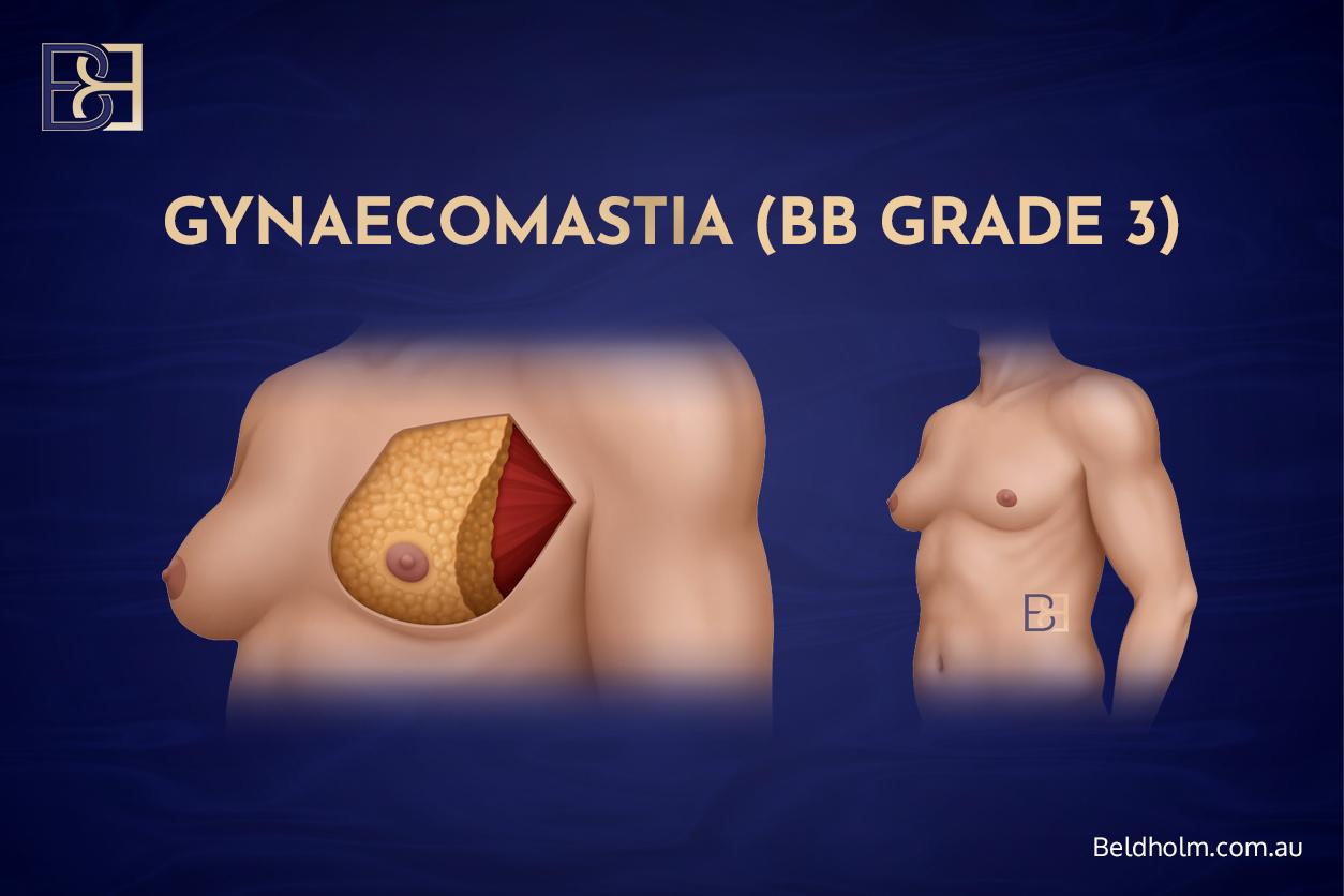
- BB grade 3: combined subcutaneous mastectomy and VASER liposuction (ultrasound-assisted lipectomy). Where there is both a glandular component and surrounding fat.
- BB grade 4: crescent lift. VASER liposuction (ultrasound-assisted lipectomy) and gland excision combined with a crescent lift, which raises the nipple by up to 2 cm by removing a crescent of skin above the areola. This suits moderate ptosis where the nipple has descended but there is still good skin elasticity, so the nipple can be raised without a full skin reduction.
- BB grade 5: mastectomy with free nipple graft. Also called torsoplasty, for severe gynaecomastia with significant excess skin and ptosis, where skin excision is needed and the nipple is repositioned as a graft, with or without VASER liposuction (ultrasound-assisted lipectomy). This is the largest of the operations and leaves the most scarring.
The landmark I use
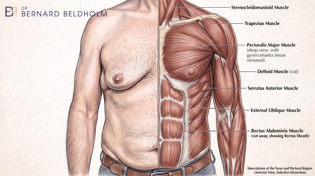
Where a grade depends on how far the nipple has descended, I measure against the inferior border of the pectoralis major muscle rather than the inframammary fold. In my experience the fold shifts and is unreliable, particularly in men who have lost weight or who have pseudo-ptosis, where the nipple has not truly dropped. The pectoralis major border is a fixed anatomical reference, so it gives a more consistent grade. This is drawn from the anatomical work behind the Gusenoff system (8).
For the full system, with each grade and the matched technique set out in detail, see: Dr Beldholm’s gynaecomastia grading system and treatment recommendations.
Why there is still no single universal system
Given how many systems exist, it is fair to ask why the surgical community has not agreed on one. There are a few reasons.
- Patients vary. Gynaecomastia ranges from a small firm disc under the nipple through to significant skin excess after major weight loss. A grading system that captures one end well often handles the other poorly.
- The mix of gland and fat differs. Some chests are mostly gland, some mostly fat, and many are a mix. A system built around gland size does not describe a fatty chest well, and the reverse is also true.
- Techniques have changed. Several of the older systems predate modern liposuction, including ultrasound-assisted liposuction. A grading system is only as current as the operations it was designed around.
- The population has changed. Two shifts stand out. More men are presenting with pseudogynaecomastia related to weight, and more men are presenting after significant weight loss with loose skin. Older systems were not built with either group in mind.
None of this means grading is pointless. It means a grade is a guide, and the right system for a given patient is the one that describes what they actually have. It also means the field keeps refining these systems as techniques and presentations change.
Pubertal gynaecomastia and younger men

A note that matters, because gynaecomastia often first appears during puberty.
Pubertal gynaecomastia is common. It tends to appear around the ages of 13 to 14 and, in the large majority of boys, it settles on its own within one to two years as hormones balance out (1,3). For that reason, surgery is not the first consideration in an adolescent. The usual and appropriate path is observation for stability, along with reassurance that this is a normal part of puberty.
Assessment becomes reasonable where the changes are established and stable rather than recent, and where they are not settling on their own. Even then, an underlying cause is looked for first, and timing is judged individually.
This article is written for adults and is not directed at people under 18. If you are a parent or a young person concerned about pubertal changes, the right first step is a GP, who can assess, reassure, and refer if needed.
Risks, recovery, and the fact that results vary
Grade influences the operation, and the operation carries risk.
Risks
Every operation on the chest carries risks, and I go through the potential complications at the initial consultation. The risks involved include:
- Haematoma, a collection of blood under the skin. This is a notable risk in gynaecomastia surgery and can require a return to theatre (11). It is one of the reasons careful control of bleeding during surgery matters, and why anticoagulants are managed carefully around the operation.
- Seroma, a collection of fluid.
- Contour irregularity and asymmetry, where the two sides do not match or the surface is uneven.
- Changes in nipple sensation, which can be temporary or lasting.
- Scarring. The higher the grade, the more this matters, which I explain below.
- Recurrence, particularly where an underlying cause has not been treated.
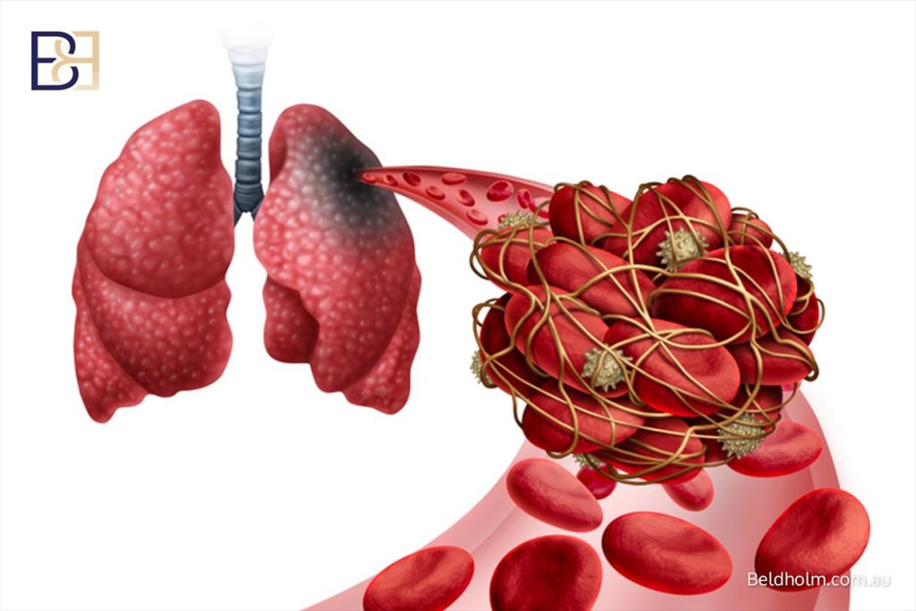
- Blood clot in the leg or lung. Deep vein thrombosis is uncommon in this operation compared with larger body contouring procedures, but it is not zero. I assess the risk for each patient and decide on prevention accordingly.
- Risks of general anaesthesia. Most of these operations are done under a general anaesthetic, and that carries its own risks, which the anaesthetist goes through separately.
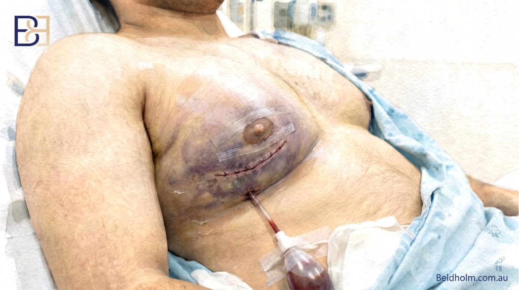
In the lower grades the work is done through small incisions. In severe cases, where skin excision is needed, the operation is larger and the potential complications increase with it.
Scars scale with grade
This is where grade has a very direct effect on what a patient should expect. In the lower grades, the incision sits at the edge of the areola and the scar is usually well hidden. In the higher grades, where skin has to be removed from the chest wall, there will be visible scars on the chest. That trade-off, accepting a scar in exchange for removing excess skin, is a genuine decision, and I talk it through with every patient before we commit to anything.
Recovery

Recovery takes time and varies between patients. Most patients need a compression garment for a period after surgery, drains are sometimes used, and return to exercise and the gym is staged rather than immediate. Pain relief is prescribed, and pain medication is usually needed for the first several days. Swelling and bruising take weeks to settle, and the healing process continues well beyond that, so the final shape is not apparent for months. I follow patients up over the first year to monitor healing.
Where a patient has had massive weight loss, I want weight to be stable before any surgical intervention. Operating at a stable weight gives a more predictable result and lowers the chance of further excess skin developing afterwards.
References
- Barros AC, Sampaio MC. Gynecomastia: physiopathology, evaluation and treatment. Sao Paulo Med J. 2012;130(3):187-97.
- Beniwal S, et al. The Burden of Anabolic Androgenic Steroid-Induced Gynecomastia. Indian J Plast Surg. 2023;56(4):338-41.
- Nydick M, Bustos J, Dale JH, Rawson RW. Gynecomastia in adolescent boys. JAMA. 1961;178(5):449-54.
- Simon BE, Hoffman S, Kahn S. Classification and surgical correction of gynecomastia. Plast Reconstr Surg. 1973;51(1):48-52.
- Cohen IK. Gynecomastia: suction lipectomy as a contemporary solution (Discussion). Plast Reconstr Surg. 1987;80(3):386.
- Rohrich RJ, Ha RY, Kenkel JM, Adams WP. Classification and management of gynecomastia: defining the role of ultrasound-assisted liposuction. Plast Reconstr Surg. 2003;111(2):909-23.
- Cordova A, Moschella F. Algorithm for clinical evaluation and surgical treatment of gynaecomastia. J Plast Reconstr Aesthet Surg. 2008;61(1):41-9.
- Gusenoff JA, Coon D, Rubin JP. Pseudogynecomastia after massive weight loss: detectability of technique, patient satisfaction, and classification. Plast Reconstr Surg. 2008;122(5):1301-11.
- Ratnam BV. A new classification and treatment protocol for gynecomastia. Aesthet Surg J. 2009;29(1):26-31.
- Punia S, Gupta A. A New Clinical Classification for Gynecomastia Management and Predictive Outcome. Indian J Plast Surg. 2023;56(4):332-7.
- Steele SR, Martin MJ, Place RJ. Gynecomastia: complications of the subcutaneous mastectomy. Am Surg. 2002;68(2):210-3.