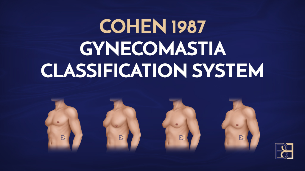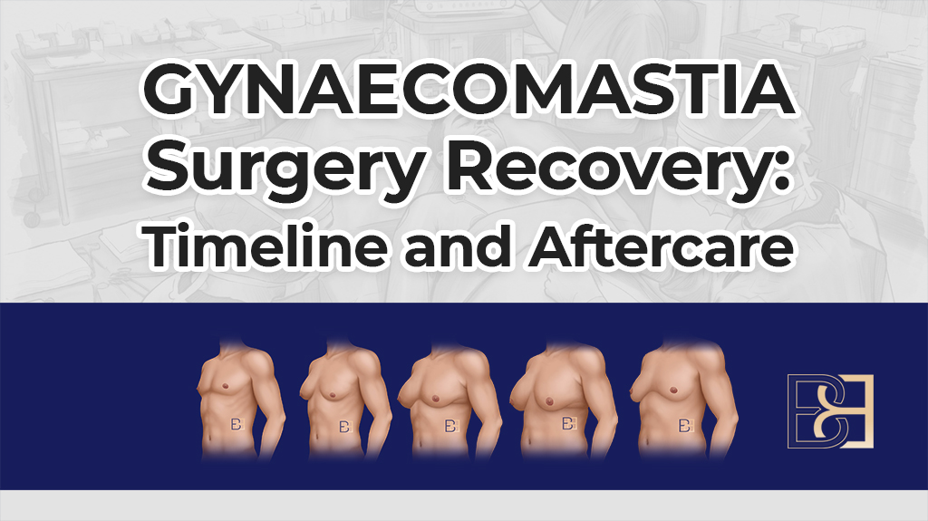
Gynaecomastia is enlarged male breast tissue. It is a recognised clinical condition with identifiable causes, and those causes matter more than most men expect when they first come to see me.
Some causes settle without any treatment at all. Some are driven by a medication or an agent that is still being taken, and stopping that agent under medical guidance changes the picture. Some point to an underlying medical condition that needs investigation before surgery is even discussed. And in a proportion of men, no cause is ever found.
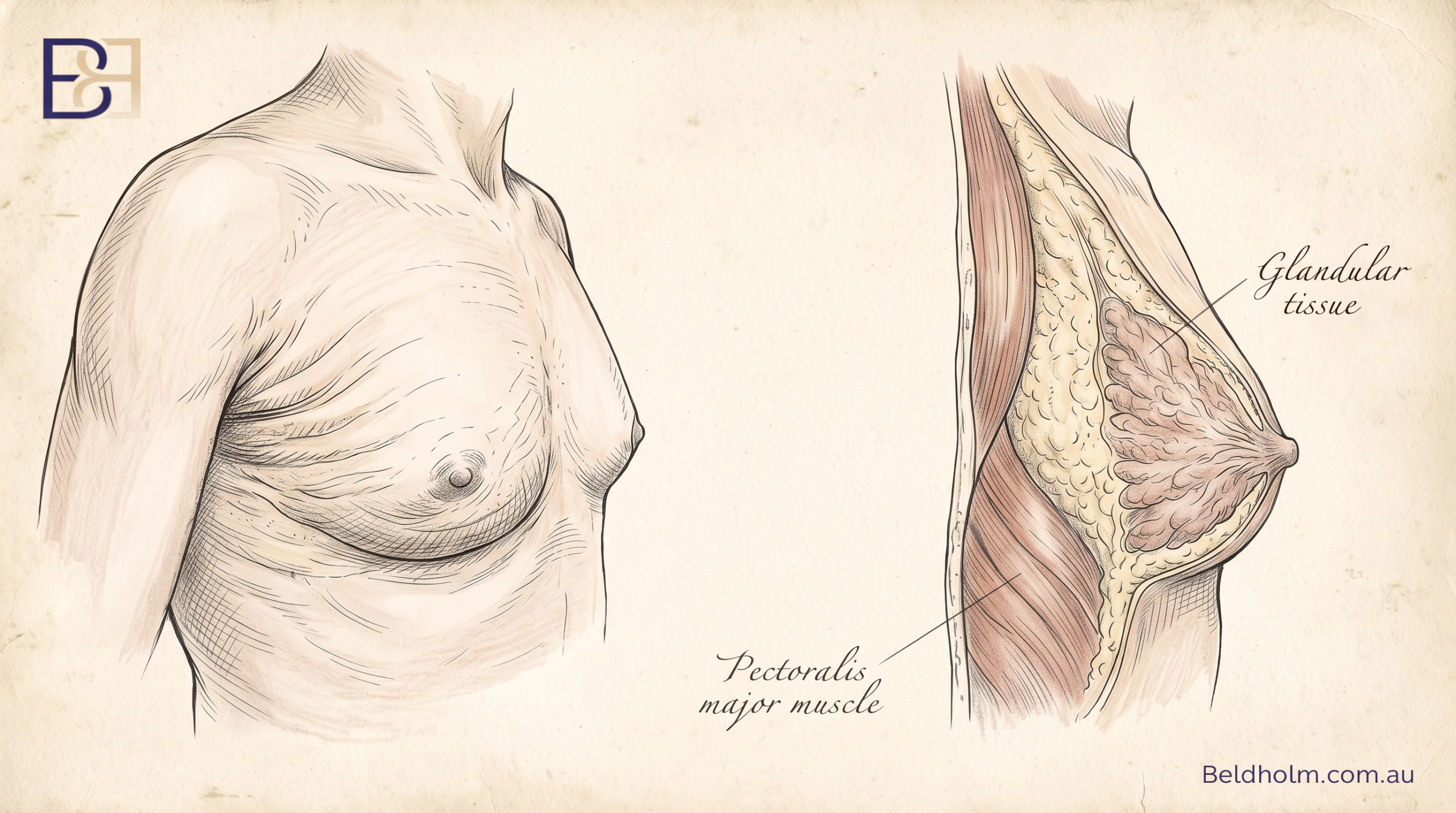

Most of the men I see have already worked out that something in the chest is firm and will not shift with training or weight loss. What they have usually not been told is why that happens, what the tissue actually is, or what it means for the operation they may or may not need.
Here I set out what gynaecomastia is and how it differs from pseudogynaecomastia, how common it is at each age, the causes I look for, the symptoms men notice, the findings that need investigation before anything else, how I assess the chest in clinic, the blood tests I order and why, how the grade of the condition maps to a specific operation, and the treatment options, including when surgery is and is not the next step.
Table of Contents
What gynaecomastia is, and what it is not
Two different tissues can make a man’s chest look fuller and give the appearance of enlarged breasts. One is glandular breast tissue. The other is fat. They look similar from the outside and they behave completely differently, which is why the first thing I do at consultation is work out which one I am dealing with.
True gynaecomastia
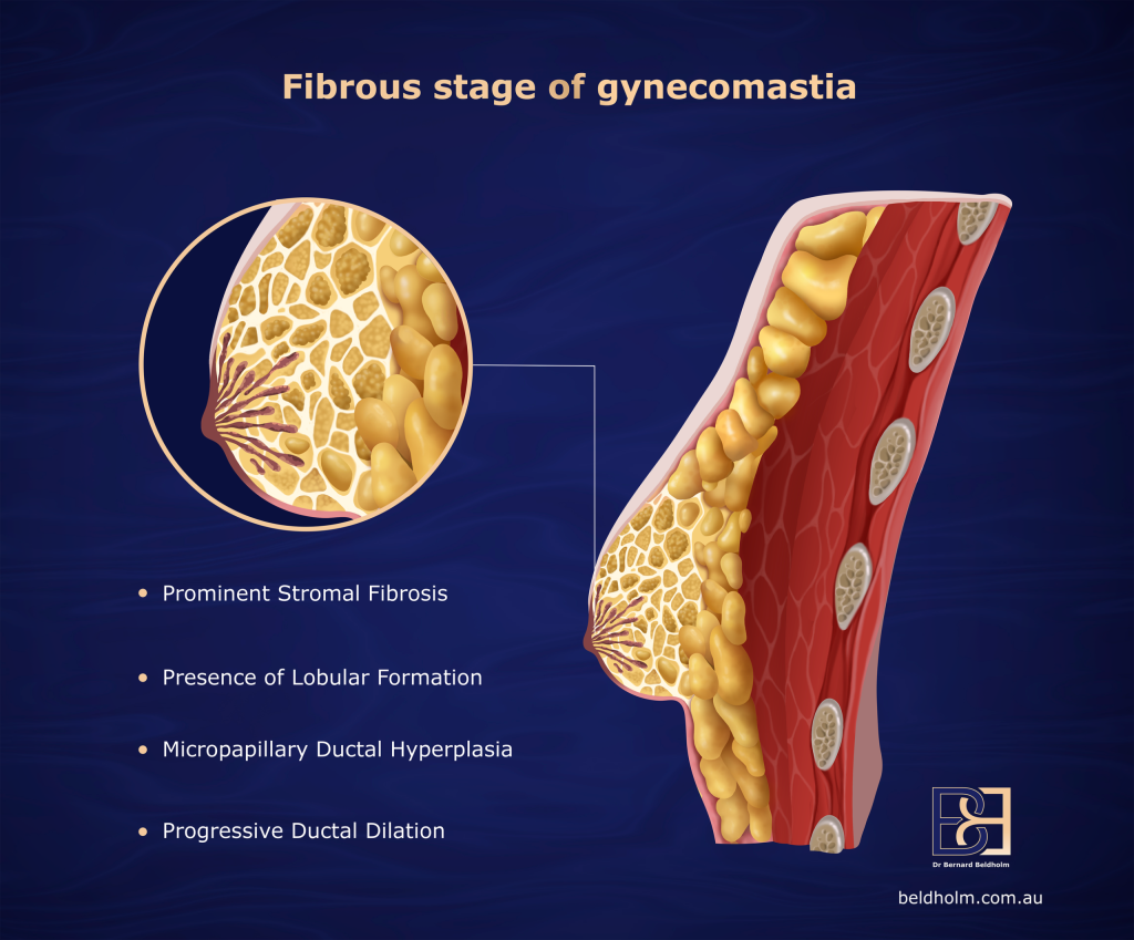
True gynaecomastia is proliferation of the glandular breast tissue itself (1). On examination it feels firm and rubbery, and it sits as a discrete disc directly behind the nipple and areola. It often extends a short distance out from the areolar edge. It can be tender, particularly when it is new. It may affect one or both breasts, and where both are involved it is frequently asymmetrical.
The important point about this tissue is that it does not behave like fat. It does not reduce with training, it does not reduce with weight loss, and men who are lean often still have a firm mound sitting under the nipple.
Pseudogynaecomastia
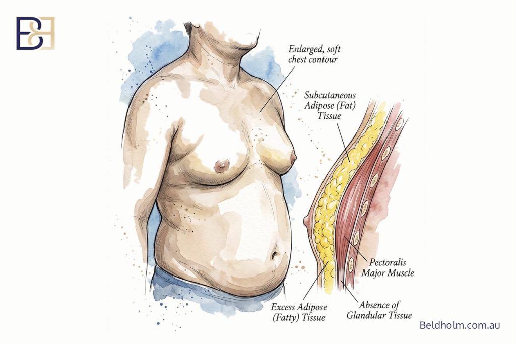
Pseudogynaecomastia, also called lipomastia, is fat sitting over the pectoralis major muscle without any glandular proliferation underneath (1). It feels soft and diffuse. There is no discrete disc to find on palpation, and the tissue blends into the fat of the surrounding chest wall rather than stopping at a defined edge.
Unlike glandular tissue, this fat does respond to weight loss.
Most men have both
In my practice the mixed presentation is the most common one. There is a fatty component across the chest and a firmer glandular disc behind the areola, in varying proportions. A man who loses a significant amount of weight will often find the chest as a whole reduces, but a firm mound remains under the nipple. That remaining mound is the gland.
One examination finding I rely on: with the arms raised above the head, fat tends to flatten out against the chest wall while the glandular disc stays prominent and becomes easier to feel and to see.
Why the distinction decides the operation
This is not an academic point. It determines what is done in the operation theatre.
- Fat is treated with VASER liposuction (ultrasound assisted lipectomy).
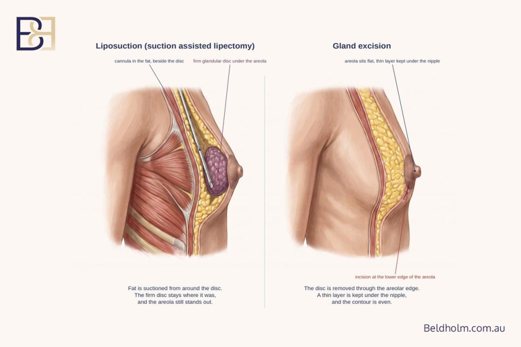
- A glandular disc is treated by excising it. Liposuction (suction assisted lipectomy) alone will not remove it.
- Where both are present, both parts of the operation are needed.
How common it is, and at what ages
Gynaecomastia is common, and it is not confined to men who are overweight. Breast tissue can enlarge at any age, and there are three points in life where it happens often enough to be considered part of normal development.
Newborn boys
Transient breast enlargement in newborn babies is caused by the high oestrogen environment of pregnancy crossing the placenta (1). It settles on its own over weeks to months and needs no treatment.
Puberty

This is the peak. Published research indicates that somewhere between 30 and 60% of boys show some degree of breast development during adolescence (1). A study of nearly 1,900 adolescent boys, with overweight boys excluded so that fat was not confused with gland, found breast tissue present in 38.7% of examinations overall, with a peak of 64.6% in boys aged 14 to 14.5 (2).
The natural history matters as much as the numbers. Mild pubertal gynaecomastia typically appears around 13 or 14 years of age, lasts 6 to 12 months, and regresses on its own in about 95% of cases (1). In the adolescent study above, the tissue was still present two years later in 27.1% of boys and three years later in only 7.7% (2).
The mechanism is a timing mismatch. Oestradiol production rises before testosterone production does, which leaves a relative excess of oestrogen at the breast tissue during that window (1). As testosterone catches up, most cases settle.
Adult men
At least a third of adult men are estimated to have some degree of glandular breast tissue (1). Most never notice it and never seek an opinion on it. The men who do come to see me are usually those in whom it is visible, firm, tender, asymmetrical, or persistent since adolescence.
Older men
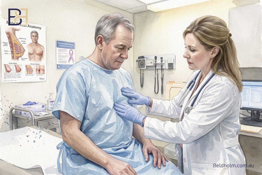
Prevalence is highest in older age, detected in up to 65% of men (1). The reasons are cumulative. Testosterone falls, sex hormone binding globulin rises so that less testosterone is free and active, body fat increases and converts more androgen to oestrogen at the tissue level, and the medications commonly used at this age can contribute (1).
What this means for an adolescent
Because most pubertal cases resolve without any treatment, surgery is not the first consideration in an adolescent. The appropriate approach is assessment to confirm what the tissue is and to exclude an underlying cause, then observation over time. Where breast tissue is still present two to three years after it appeared, and the boy has otherwise completed puberty, it is less likely to settle on its own and assessment for treatment becomes reasonable. That decision is made with the family, the GP, and where indicated a paediatric endocrinologist.
The underlying mechanism
Male breast tissue carries receptors for both oestrogen and androgen. Oestrogen stimulates the breast ducts to proliferate. Androgen does the opposite and holds that process in check (1). Gynaecomastia develops when the balance between the two shifts at the level of the breast tissue.
That shift can happen from either direction.
More oestrogen effect, through increased secretion by the testes or adrenal glands, increased peripheral conversion of androgen precursors into oestrogen, reduced breakdown of oestrogen, exposure to oestrogen or oestrogen-like substances from outside the body, or a drug that displaces more oestrogen than androgen from sex hormone binding globulin (1).
Less androgen effect, through reduced testosterone production by the testes, increased binding of testosterone by sex hormone binding globulin so that less is free and active, altered androgen metabolism, a drug that blocks androgen at the receptor, or a defect in the androgen receptor itself (1).
Why fat is part of the mechanism, not just the appearance
In men, only a small proportion of circulating oestrogen comes directly from the testes. Around 80% is produced by peripheral conversion of androstenedione and testosterone, driven by the enzyme aromatase. That conversion happens mainly in subcutaneous fat, including the fat within the chest itself, and aromatase activity increases both with age and with rising body mass index (1).
This is a point I make often at consultation. Chest fat is not only a cosmetic component of the problem. It is metabolically active tissue producing oestrogen next to the gland it stimulates. It is one reason weight and gynaecomastia are linked, and one reason weight stability matters before surgery in a man with a significant fatty component.
Why blood tests are often normal
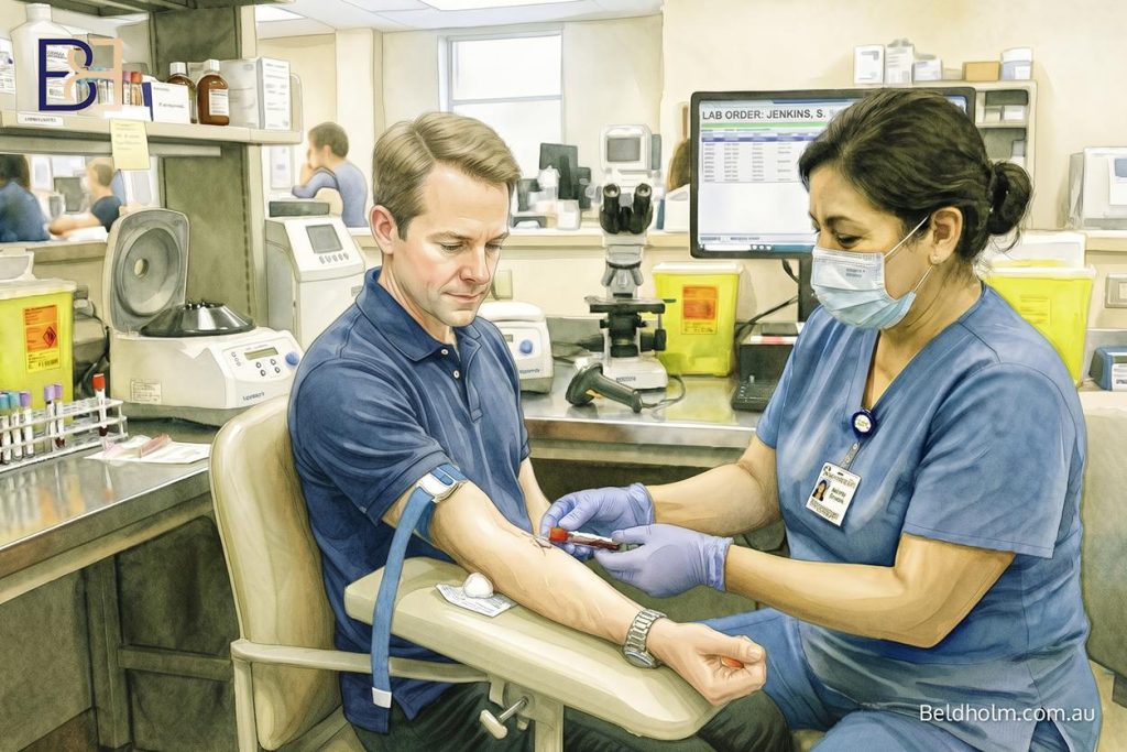
Because the imbalance operates locally at the breast tissue, and because the amounts involved are small relative to circulating hormone levels, many men with clear gynaecomastia have an entirely normal endocrine panel. A normal set of results does not mean the diagnosis is wrong. It means no systemic cause has been identified, which is useful information in itself.
Early tissue and established tissue behave differently
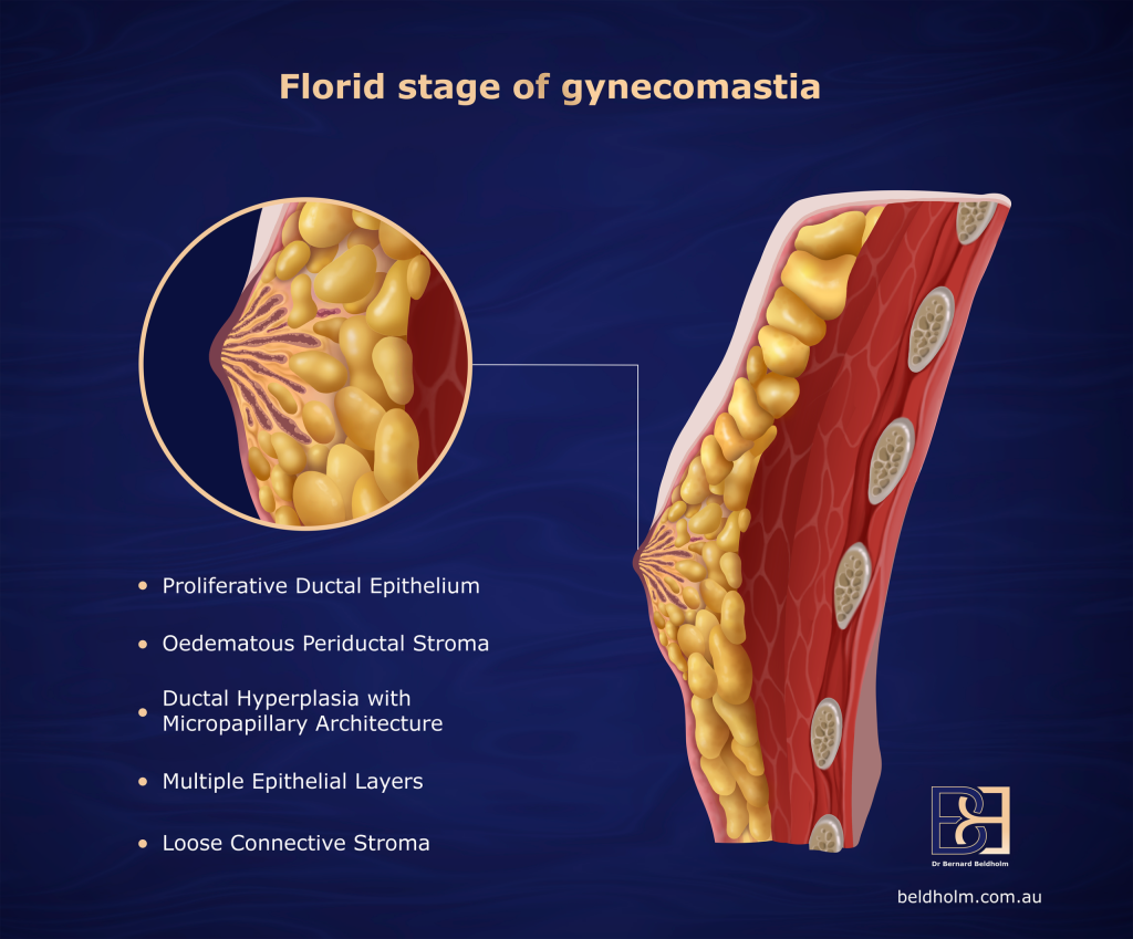
Under the microscope, gynaecomastia is proliferation of the breast ductules within fibroconnective stroma. In the early stage, called the florid phase, there is extensive ductal hyperplasia. Over time the glandular elements become less prominent and fibrosis takes over, which is called the fibrous phase (1).
This has two consequences that matter to the man sitting in front of me.
- Medication has a chance of working only in the early proliferative phase, before the glandular structure has been replaced by fibrosis (1). Once tissue is fibrous, medical treatment is not going to reduce it.
- Tissue that has been present for more than a year, which is most of what I operate on, is fibrous rather than florid (3). That is the firm disc men feel, and it is why it does not respond to training, to weight loss, or to liposuction (suction assisted lipectomy) alone.
What causes gynaecomastia
The recognised causes of gynaecomastia fall into a small number of groups: physiological change at particular stages of life, medications and other agents, endocrine dysfunction, other health conditions, tumours, and idiopathic cases where no cause is ever identified (1). Some of these are risk factors that stack together in the same man rather than acting alone.
Working through these in order is the part of the consultation that most men have not had done before.
Physiological causes
Puberty and ageing account for a large share of cases, and both were covered above. In an older man the drivers stack: falling testosterone, rising sex hormone binding globulin, increasing body fat and therefore increasing aromatase activity, and a growing list of medications (1).
Medications

Medications are one of the largest identifiable groups, estimated to account for roughly 20 to 25% of cases (1, 4). The list is long and it crosses most therapeutic classes.
Class | Agents reported to cause gynaecomastia |
|---|---|
Anti-androgens | bicalutamide, cyproterone, flutamide, finasteride, spironolactone |
Antibiotics and antifungals | isoniazid, ketoconazole, metronidazole |
Antihypertensives, including calcium channel blockers | amlodipine, captopril, enalapril, nifedipine, verapamil, reserpine |
Chemotherapeutic agents | cyclophosphamide, methotrexate |
Gastrointestinal agents | cimetidine, ranitidine, omeprazole, metoclopramide |
Hormones | androgens, anabolic steroids, oestrogens, growth hormone |
Psychiatric agents | diazepam, haloperidol, phenothiazines, tricyclic antidepressants |
Other | amiodarone, antiretrovirals, digitalis, domperidone, statins, theophylline |
Adapted from published data on drug-induced gynaecomastia (1).
Two points about this table. The first is that appearing on it does not mean a drug caused a particular man’s gynaecomastia. Many of these agents are widely prescribed and the association is uncommon. The second is more important: no one should stop a prescribed medication because they have read this list. Several of these drugs are treating conditions where stopping without supervision carries greater risk than the breast tissue does. Where a medication looks like a plausible driver, the conversation is between the patient, the GP and the prescribing specialist, and it is about whether an alternative agent exists.
Alcohol and cannabis are also reported causes, both through effects on the oestrogen to androgen balance (1).
Anabolic androgenic steroids
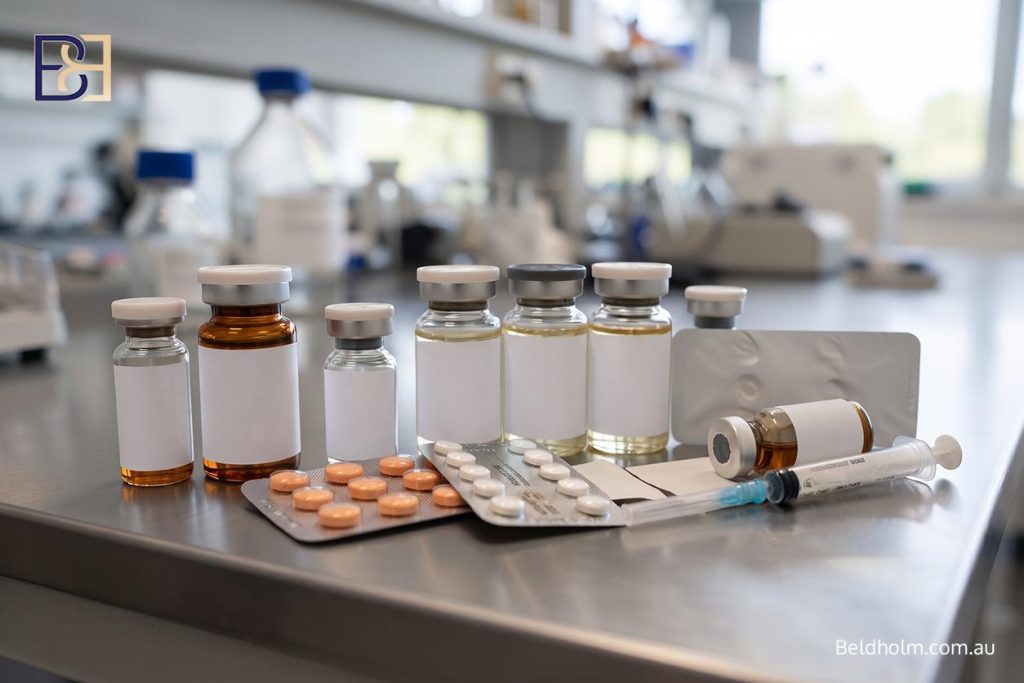
This is a significant group in the men I operate on, and it is the group most often not disclosed at the first consultation.
Anabolic androgenic steroids are aromatised to oestrogen within the body, which produces exactly the imbalance described earlier. A prospective study makes the disclosure problem clear. Of 74 men treated for gynaecomastia, only 4% gave a history of steroid use before surgery. When the same men were asked again three months after surgery, the figure was 39% (4). In that series, men who had used steroids to build muscle mass had a higher body mass index, were more often bodybuilders, and had less fat and proportionally more gland at operation (4).
It matters for three reasons.
- Where the agent is stopped, some cases regress without any operation. Where the tissue has been present beyond about a year, dense fibrous tissue predominates and complete regression becomes unlikely (4).
- The operation is different. These chests tend to carry proportionally more glandular tissue and less fat, which changes the technique and the dissection required.
- Continuing to use the agent after surgery means the driver is still active, which is the single clearest route to recurrence.
Endocrine dysfunction
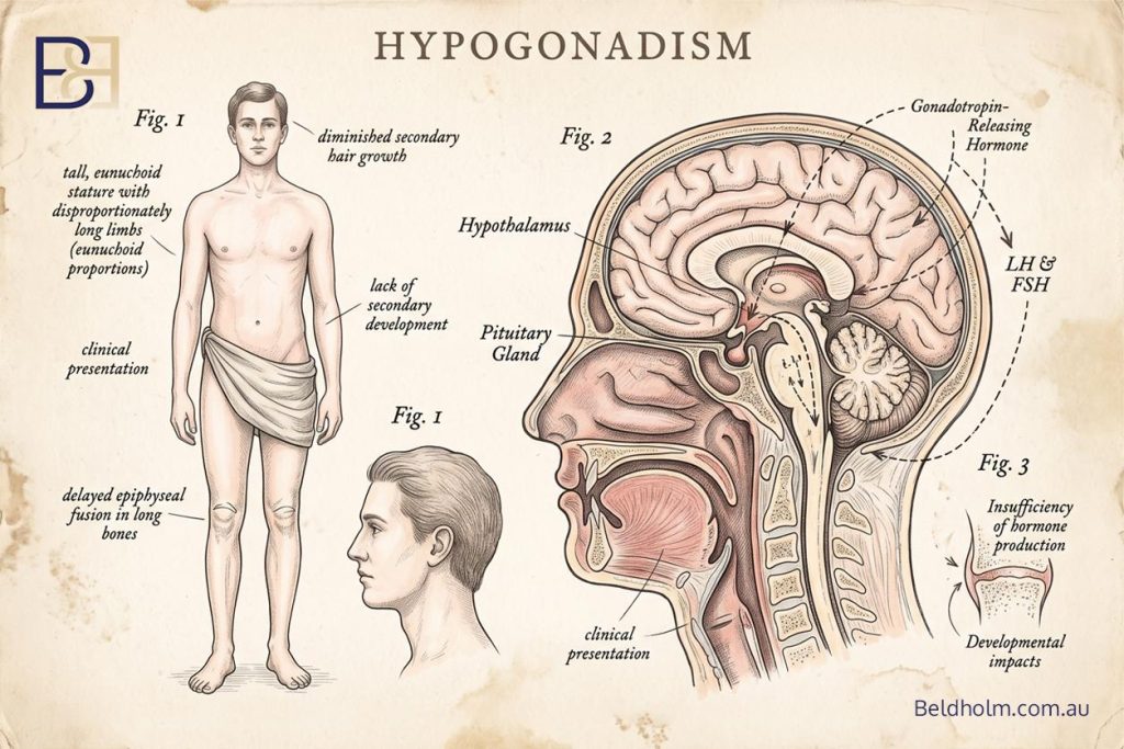
Hypogonadism, whether primary or secondary, is a recognised cause. It lowers testosterone levels, and in some men the testes are small, a finding described as testicular atrophy. Published estimates attribute around 8% of cases to primary hypogonadism and around 2% to secondary hypogonadism (1). Hyperthyroidism accounts for around 1.5%, through a rise in sex hormone binding globulin (1). Obesity contributes through aromatase activity in fat, and refeeding after a period of malnutrition or significant weight loss can trigger it as hormonal function recovers (1).
Non-endocrine disease
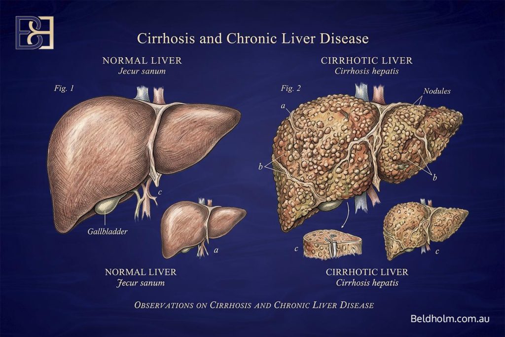
Cirrhosis and chronic liver disease account for around 8% of cases, chronic renal failure, also called kidney failure, around 1%, and gynaecomastia occurs in 2 to 3% of men with HIV, related either to lipodystrophy or to antiretroviral therapy (1).
Tumours
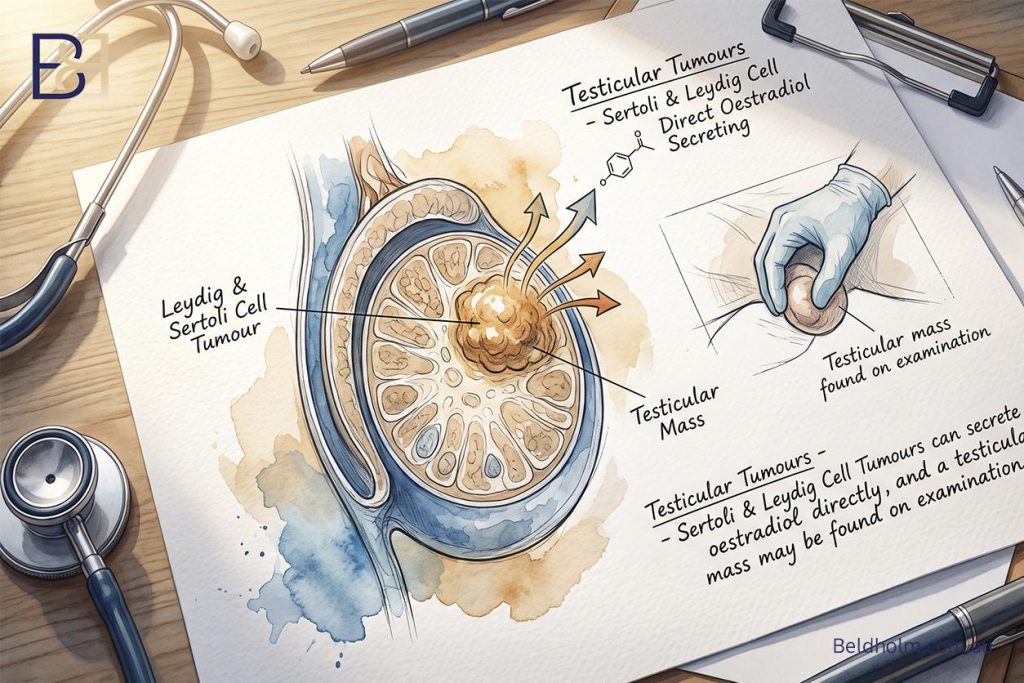
This group is uncommon.
- Testicular tumours account for around 3% of cases. Sertoli and Leydig cell tumours can secrete oestradiol directly, and a testicular mass may be found on examination (1).
- Germ cell tumours, including choriocarcinoma, produce hCG, which stimulates the testis to secrete oestradiol. Some of these are palpable and some are found only on ultrasound (1).
- hCG-secreting tumours elsewhere, including in the lung, liver, stomach and kidney, can do the same (1).
- Prolactin-secreting adenomas of the pituitary gland can induce gynaecomastia (1).
- Adrenocortical tumours secrete oestrogens and steroid precursors directly (1).
This is a short list of rare diagnoses, and it is why beta-hCG, prolactin and the rest of the endocrine panel are ordered rather than skipped.
Idiopathic
In a substantial proportion of men, every test comes back normal and no cause is found. It does not change what can be done surgically. It does mean there is no ongoing driver to treat first, which in practice simplifies the timing decision.
Symptoms and what men notice
Most men do not present because of a symptom in the medical sense. They present because of something they can feel or see that has not changed over time. The clinical picture is usually consistent.
A firm lump behind the nipple
This is the most common description I hear. Something firm sits directly under the nipple and areola, it feels like a disc or a button rather than soft tissue, and it moves with the skin rather than being fixed to the muscle underneath. It is concentric with the nipple, which is one of the features that separates it from other lesions.
Tenderness
Tenderness is common, particularly in the early stage while the tissue is still in the florid phase. It can be a dull ache, sensitivity to pressure from a seatbelt or a backpack strap, or discomfort during exercise. In one series, pain was reported by 43% of men presenting for treatment (4).
Tenderness usually settles as the tissue becomes fibrous over the following months. Its absence does not mean the tissue has resolved. It commonly means the tissue has matured.
Prominence of the nipple and areola
The areola can appear fuller or more domed, and the nipple can sit forward of the chest wall rather than flat against it. This is the tissue immediately behind the nipple pushing the areola outwards. Some men describe it as the nipple showing through clothing, particularly in fitted shirts or after training.
Asymmetry, and one side only
Gynaecomastia is clinically bilateral in more than half of men in some series (1). Where it is present on both sides, the two sides are frequently different in size. In the large adolescent series, it was unilateral in 23.3% of cases, more often on the right (2).
Asymmetry is normal for this condition and it is expected. A difference between the two sides is not in itself a concerning finding. What matters is the pattern, and I cover the findings that do need investigation in the next part.
Nipple discharge
Discharge from the nipple is very uncommon in gynaecomastia (1). Where it does occur it is not attributed to gynaecomastia without investigation.
What men also notice: it does not respond to what works elsewhere
The description I hear most often is a man who has lost weight, or trained consistently, or both, and has seen the rest of his body change while the chest has not. In some cases the tissue looks more obvious after weight loss, because the fat that was blending it into the surrounding chest has gone and the disc underneath has not.
That pattern is not a training failure. It is the difference between fat and glandular tissue.
Findings that need investigation before anything else
Breast cancer in men is uncommon, but it exists, and it presents in the same anatomical area as gynaecomastia. Excluding it is part of the assessment rather than an optional extra. There are also several benign lesions that can be mistaken for gynaecomastia, including lipomas, dermoid cysts, neurofibromas, lymphangiomas and old post-traumatic haematomas (1).
The pattern of the findings is what separates them.
How the two present differently
Gynaecomastia is felt as a disc of firm tissue that is concentric with the nipple and areola, meaning it sits centred behind the nipple and extends outwards evenly (1).
Breast carcinoma in men usually presents as a unilateral, hard, irregular mass sitting outside the areola rather than centred on it, and it may be accompanied by dimpling of the skin, retraction of the nipple, or enlarged lymph nodes in the armpit (1).
Findings that need a GP assessment before a surgical opinion
- A hard or irregular mass, as opposed to a firm rubbery disc
- A lump that sits off to one side rather than centred behind the nipple
- A mass that feels fixed to the skin or to the chest wall underneath
- Dimpling, puckering or tethering of the overlying skin
- Nipple retraction, or a change in the direction the nipple points
- Ulceration or a persistent change in the skin of the areola
- Nipple discharge, particularly if it is blood-stained
- Enlarged lymph nodes in the armpit
- Rapid enlargement on one side, especially in an adult with no obvious cause
- Any breast change accompanied by a lump in a testis
The last one matters. A testicular tumour secreting oestradiol or hCG can present with breast enlargement, and the testicular examination is part of the workup for that reason (1).
Two groups warrant a lower threshold for imaging: men who have family members with breast cancer, and men with Klinefelter syndrome (1).
What investigation involves
Where there is any suspicion of malignancy, mammography is the primary imaging method. As a predictor of malignancy in this setting it has a reported sensitivity of 90% and a specificity of 92% (1). Ultrasound is widely used and gynaecomastia has recognised sonographic patterns, typically a hypoechoic mass behind the areola (1). Where imaging and examination together cannot separate gynaecomastia from carcinoma, a percutaneous biopsy is performed (1).
Tissue is sent for histopathology
Every specimen I excise goes to pathology. It is because the only way to be certain what tissue was removed is to have it examined, and occasional unexpected diagnoses are reported in published surgical series (3).
If any of the findings listed above apply, see a GP first. That pathway is a different one to a surgical consultation and it takes priority over it.
How I assess gynaecomastia in clinic

The consultation has a fixed structure. I am answering four questions: what is the tissue, what is driving it, does anything need excluding first, and what would the operation actually be.
History
The history does most of the diagnostic work. I ask about:
- When it appeared, how quickly, and whether it has changed since
- Whether it is tender, and whether it was more tender early on
- Whether it is one side or both, and whether the two sides differ
- Every prescribed medication, including agents started in the months before it appeared
- Supplements, over the counter preparations, alcohol intake and recreational drug use
- Anabolic androgenic steroid use, past and current, and whether use is intended to continue
- Weight history, including the highest and current weight, any weight loss medication use, and whether weight has been stable
- Symptoms suggesting low testosterone levels, including reduced libido, erectile dysfunction, fatigue and reduced body hair
- Symptoms suggesting thyroid, liver or kidney disease
- Family history of breast cancer, and any history of testicular problems or undescended testis
I ask about anabolic androgenic steroids as a routine part of the medication history, in the same way I ask about anything else that has been taken. It is a clinical question rather than a judgement, and the answer does not change the care given. Many men have never had the link between the agent and the breast tissue explained to them, so it does not occur to them that it is relevant. Published data show reported rates rise substantially when men are asked again later in the treatment pathway, which is why I explain why I am asking rather than simply asking (4).
Examination
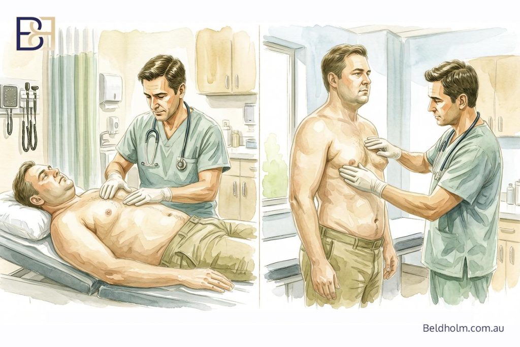
I examine the chest with the man lying down and again standing, because the two positions show different things.
Separating gland from fat. With the patient supine and hands behind the head, I palpate from the outer chest wall inwards towards the nipple using thumb and forefinger. Glandular tissue announces itself as a firm, mobile, rubbery disc with a definable edge, concentric with the areola. Fat has no edge and blends continuously into the surrounding chest (1).
The arms raised manoeuvre. Raising the arms above the head is part of the same assessment, since it changes how fat and gland behave and helps confirm which one is present.
Nipple position, standing. Ptosis is assessed with the man standing, measured at the centre of the nipple. The reference landmark is the inferior border of the pectoralis major muscle, not the inframammary fold, because the fold descends with the skin envelope while the muscle border does not. Whether the nipple sits above that line, within about 2 cm below it, or more than 2 cm below it is the single measurement that separates the higher grades from each other.
Skin quality. I assess three things: the extent of stretch marks, how the skin recoils when pinched and released, and how loose it sits at rest. These give a skin elasticity grade from 1 to 5. Skin that springs back will retract on its own once tissue is removed. Skin that does not will need to be excised, and at the far end of the range the nipple has to be repositioned as a graft. Skin quality is the variable that most often decides whether an operation is staged.
The rest of the examination. Both testes are examined. I also look for signs of liver disease, thyroid disease and hypogonadism, since these change what is investigated before surgery is discussed.
Photographs and grading
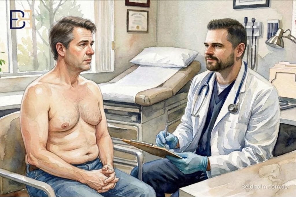
Standardised photographs are taken at consultation. The findings then map onto a grade, which is what determines the operation. That process is set out in the BB grading system and in my comparison of the published gynaecomastia grading systems.
The diagnostic workup
Blood tests in gynaecomastia have one purpose: to identify or exclude an underlying cause before any decision about surgery is made. They are ordered as the history and examination indicate, not as a fixed panel for every man. A boy of fifteen with tender breast tissue that appeared six months ago and a normal examination needs very little. An adult with rapid unilateral enlargement, or a firm chest with no obvious explanation, needs the full workup.
The endocrine panel
Test | What it helps identify |
|---|---|
Total testosterone | Hypogonadism of any cause. Measured on a morning sample, since levels follow a daily rhythm |
Luteinising hormone (LH) and follicle stimulating hormone (FSH) | Whether low testosterone is coming from the testis or from the pituitary. Low testosterone with high LH and FSH points to the testis. Low testosterone with low or normal LH and FSH points higher up |
Oestradiol | An oestrogen-secreting source, or increased conversion of androgen to oestrogen |
Sex hormone binding globulin (SHBG) | How much testosterone is actually free and active. Rises in hyperthyroidism and in liver disease |
Prolactin | A prolactin-secreting pituitary adenoma (1) |
Beta-hCG | An hCG-secreting tumour |
Thyroid function tests | Hyperthyroidism, which accounts for around 1.5% of cases (1) |
Liver function tests | Cirrhosis and chronic liver disease, around 8% of cases (1) |
Urea, electrolytes and creatinine | Chronic renal failure (1) |
Why beta-hCG is on that list
This is the test men ask about, because hCG is familiar as a pregnancy marker. In this setting it has nothing to do with pregnancy.
Germ cell tumours of the testis, including choriocarcinoma, secrete hCG, which stimulates the testis to produce oestradiol and can present as breast enlargement. Tumours elsewhere, including in the lung, liver, stomach and kidney, can secrete hCG and do the same thing (1). Some of these tumours are palpable on examination and some are not.
Imaging and referral
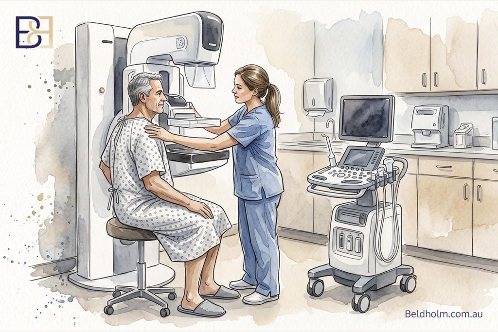
Imaging follows the findings rather than being routine.
- Testicular ultrasound where the testicular examination is abnormal, or where oestradiol or beta-hCG is raised.
- Breast imaging, mammography and ultrasound, where the examination raises any suspicion of malignancy.
- Pituitary imaging where prolactin is elevated and other causes have been excluded.
- Further imaging where beta-hCG is raised but the testes appear normal, since the source may be elsewhere.
- Endocrinology referral where an endocrine abnormality is identified, before surgery is discussed further.
Where the workup identifies a cause, treating that cause comes first. In some men that is enough on its own, particularly early in the course.
Pre-operative bloods are a separate set of tests

The endocrine workup asks what caused this. Pre-operative bloods ask whether a man is fit for an anaesthetic and an operation. They are ordered once surgery is actually planned, not at the first consultation.
For a day case gynaecomastia operation, that means a standard pre-operative set: full blood count, coagulation screen, liver function, electrolytes, glucose and HbA1c.
A nutritional panel is not routine for gynaecomastia. I add it where there is a weight loss history, because gaps that develop during significant weight loss affect wound healing and are worth correcting before an operation rather than after it.
Results are copied to the GP, who holds the long term management of anything the workup identifies.
Grading, and why it decides the operation
Several classification systems for gynaecomastia have been published over the last sixty years. Most of them describe how much tissue is present and how much the skin has stretched. What they generally stop short of doing is stating which operation follows from which finding.
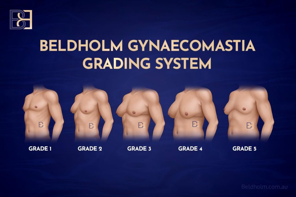
I developed my own grading system for that reason. It has five grades, and each grade maps to one operation. When I tell a man at consultation which grade he is, I am telling him what the surgical treatment is.
The five grades
Grade | What is found on examination | The operation |
|---|---|---|
BB grade 1 | Glandular disc only, confined within the areola. No significant fatty component | Gland excision through an infra-areolar incision. No liposuction (suction assisted lipectomy) |
BB grade 2 | Fat predominant, pseudogynaecomastia. No firm glandular disc | VASER liposuction (ultrasound assisted lipectomy) alone |
BB grade 3 | Both a glandular disc and a fatty component, with no ptosis | VASER liposuction (ultrasound assisted lipectomy) combined with gland excision |
BB grade 4 | Ptosis with skin that still has elasticity. Nipple sits up to about 2 cm below the inferior border of pectoralis major | Crescent lift, up to about 2 cm, with VASER liposuction (ultrasound assisted lipectomy) and gland excision |
BB grade 5 | Ptosis where the skin will not retract, or the nipple sits more than 2 cm below the inferior border of pectoralis major | Mastectomy with free nipple-areola graft, with or without VASER liposuction (ultrasound assisted lipectomy) |
Two things the table does not show
Skin quality sits across the whole system. The grade describes what tissue is present and where the nipple sits. Skin elasticity determines whether the skin will retract once that tissue is removed or whether it has to be excised. Two men with the nipple in the same position can end up with different operations because their skin behaves differently.
The middle of the skin range is a decision point rather than an answer. Where skin elasticity is borderline, my approach is to perform the BB grade 4 operation first, reassess the skin at six to twelve months, and convert to a BB grade 5 operation only if the skin has not retracted. That sequence avoids removing skin that would have tightened on its own, at the cost of a possible second operation. Which of those two a man prefers is part of the discussion.
Where each grade is covered in detail
Book your appointment online now
Each grade has its own page setting out the operation, the recovery and the complications specific to it.
- BB grade 1 gynaecomastia surgery
- BB grade 2 gynaecomastia surgery
- BB grade 3 gynaecomastia surgery
- BB grade 4 gynaecomastia surgery
- BB grade 5 gynaecomastia surgery
The system itself, including how it compares with the published classifications, is set out in the BB grading system article.
The grade is assigned at examination. It cannot be assigned from a photograph, from a description over the phone, or by a man reading this page and matching himself to a row in the table. Skin quality in particular has to be felt.
Non-surgical management and observation
Surgery is one of several treatment options, not the automatic one. For a proportion of men the right next step is to do something other than operate, at least to begin with. There are four situations where that applies.
Observation in pubertal cases

Most pubertal gynaecomastia settles on its own, so in an adolescent the usual approach is to confirm what the tissue is, exclude an underlying cause, and then wait. Reviewing the chest over time tells you whether it is regressing, staying the same, or still enlarging. Where it is still present two to three years after it appeared, and puberty is otherwise complete, spontaneous regression becomes less likely and assessment for treatment becomes reasonable. That is a decision made with the family, the GP, and a paediatric endocrinologist where indicated.
Treating the underlying cause
Where the workup identifies a cause, treating that cause comes first, because operating around an active driver leaves the driver in place. Hyperthyroidism is managed. Liver or kidney disease is managed. An endocrine abnormality is referred and treated. In some men, dealing with the cause reduces the breast tissue on its own and no operation is needed.
Reviewing a causative medication or agent
Where a prescribed medication is a plausible driver, the question is whether an alternative exists. That conversation is between the patient, the GP and the prescriber. It is never a matter of a patient stopping a prescribed medication on their own, because several of the drugs involved are treating conditions where stopping without supervision carries greater risk than the breast tissue does.
Where anabolic androgenic steroids are the driver, the position is clearer. These agents are aromatised to oestrogen, which is the imbalance that produces the breast tissue. Published data show that where the agent is stopped early, some cases regress without any operation, and that men who stop and are managed conservatively can avoid surgery altogether (4). The important qualifier is timing. Once the tissue has been present beyond about a year it is largely fibrous, and fibrous tissue does not regress. At that point stopping the agent protects against the tissue getting worse and against recurrence after surgery, but it will not clear a disc that has already formed.
Weight stability where there is a fatty component
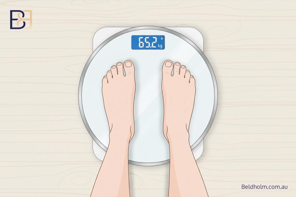
Where a significant part of the fullness is fat, weight matters. Fat in the chest is metabolically active and contributes to the hormonal imbalance, so a man who is actively losing weight is aiming at a moving target. Planning surgery once weight is stable gives a more predictable result and avoids operating on a chest that is still changing. This applies to the fatty component. It does not apply to an established glandular disc, which will not shift with weight loss whatever the number on the scales.
Medication for gynaecomastia itself
Medication can reduce gynaecomastia, but only within a narrow window. It has a chance of working in the early proliferative phase, before the glandular tissue has been replaced by fibrosis (1). Once the tissue is fibrous, which is the case for most established gynaecomastia, medical treatment is not going to reduce it (1).
The agents that have been studied are the selective oestrogen receptor modulators, mainly tamoxifen, and the aromatase inhibitor anastrozole. Tamoxifen has the better evidence of the two. A series of idiopathic cases treated with tamoxifen reported regression in around 80% (1), and tamoxifen has been shown to reduce the gynaecomastia and breast pain caused by certain prostate cancer treatments (1). Anastrozole has weaker support. In a randomised controlled trial in pubertal boys it was no more effective than placebo (1).
Several points matter here. None of these drugs is approved specifically for gynaecomastia, the overall quality of evidence is low, and there is no agreed drug of choice or duration of treatment (1). Prescribing them is a decision for a treating doctor after a discussion of the evidence and the potential harms, and it is not something I initiate as a surgeon. Where medication is appropriate, it belongs early in the course, and it belongs with the GP or an endocrinologist rather than in a surgical clinic.
What non-surgical management does not do
None of the above changes a firm, established, fibrous disc. Observation, treating the cause and stopping a driver are the right first steps, and for some men they are enough. If a man has a firm glandular disc that has been there for years, these steps still have a role. They manage the underlying cause and they lower the chance the tissue gets worse or comes back. What they cannot do is remove the disc that has already formed. Surgery is the only thing that removes it.
When surgery is considered, and when it is not
By this point the two questions that decide timing have already been answered: what is driving the gynaecomastia, and is the tissue the kind that will respond to anything other than surgery. Surgery comes into consideration once those are settled.
When surgery is considered
I consider surgery once these conditions are met.
- An underlying cause has been excluded or managed. The workup is done, and where it found something, that has been treated. Operating with an active, treatable cause still running leaves the driver in place.
- A causative agent has been stopped where appropriate. Where a medication or an anabolic androgenic steroid was driving the tissue, it has been reviewed and, where appropriate to do so under medical guidance, stopped.
- The condition is stable. The tissue is established rather than recent, and it is not still enlarging. In pubertal cases this means it has been observed long enough to know it is not going to settle on its own.
- Weight is stable, where there is a significant fatty component. The chest is not still changing from ongoing weight loss.
- The tissue will not respond to anything else. It is a mature glandular disc, not early proliferative tissue that medication might reduce.
- The man understands the operation. He understands what it involves, the recovery, the risks including haematoma, and that the outcome differs from one person to the next.
When those things line up, surgery is a reasonable option to discuss.
When surgery is not the first step
Equally, there are situations where operating is not the right next move.
- Recent pubertal gynaecomastia. It is common, it frequently settles on its own, and in an adolescent it is observed rather than operated on early.
- A cause that has not been investigated. Where the workup has not been done, it is done first. Operating without it risks missing something that needed treating in its own right.
- A causative agent still in use that has not been reviewed. Where a medication or an anabolic androgenic steroid is still driving the tissue and has not been looked at, that review comes first.
- Weight that is not yet stable, in a man whose fullness is substantially fat. Surgery is planned once weight has settled.
In several of them the right first step reduces the tissue anyway, and where it does not, surgery is still there once the groundwork is done.
The decision is made at consultation, for the individual
Whether surgery is appropriate, and which operation, is a clinical judgement made for the man in front of me. It follows the history, the examination that separates gland from fat, and the workup. It cannot be made from a photograph or over the phone, and it cannot be made before assessment.
This is also why timing is never a fixed number of weeks or months. It depends on the cause, on how long the tissue has been present, on whether an agent has been stopped, and on whether weight is stable.
Recurrence, and why the cause still matters after surgery
Surgery removes the tissue that is there. It does not change what caused that tissue to form. This is the point I want a man to understand before he books an operation.
The clearest example is an ongoing agent
Anabolic androgenic steroids are the most direct illustration. If the agent is still being used after surgery, the hormonal imbalance that caused the tissue is still present, and that is the single clearest route back to enlarged tissue. Continuing to aromatise an androgen to oestrogen after an operation undoes part of what the operation achieved.
Weight is the other common one
Where a significant part of the fullness was fat, and a man regains substantial weight afterwards, the chest can enlarge again. In practice this is weight-related change rather than the original glandular tissue regrowing. It is one of the reasons weight stability is part of the timing decision in the first place.
Recurrence of the gland itself is uncommon when the operation is complete
Where the glandular disc is properly excised and no ongoing driver is present, the same gland regrowing is uncommon. The disc has been removed and there is no active process reforming it.
The risks, in brief
This is surgery, and surgery carries risk. Each grade has its own complications page, because the risks of an operation for a fat-predominant chest and an operation involving a nipple graft are not the same. What follows is the short overview.
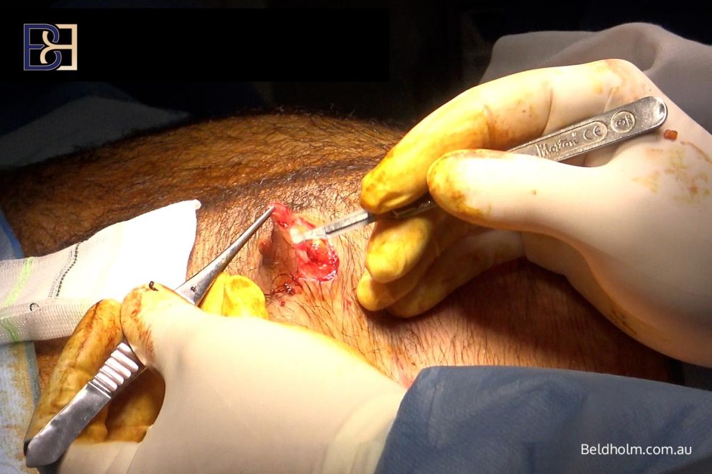
The risk to understand first is haematoma, a collection of blood under the skin after surgery. It is consistently the most common complication in this operation, and it is more likely where the glandular component is large, because glandular tissue bleeds more than fat (5). It is the reason for careful control of bleeding during surgery, drains in the higher grades, a compression garment afterwards, and stopping anything that thins the blood beforehand under medical guidance.
The other recognised complications are seroma, contour irregularity, under-correction or over-correction, asymmetry, a change in nipple sensation that is usually temporary but can be lasting, scarring, infection, and the general risks of an anaesthetic. Reported complication rates across the literature vary widely with the grade, the technique and how complications are defined (6). The pattern is consistent though: the more tissue and skin involved, the higher the rate. That is one more reason the operation is matched to the grade.
Some men need a second procedure, to even out a contour, revise a scar, or remove residual tissue. In the higher grades a planned second stage is sometimes part of the approach from the outset rather than a complication.
The risk and recovery detail for each operation is set out on the individual grade pages.
Dr Beldholm’s conclusion

If there is one thing I want a man to take from this page, it is that gynaecomastia is not one thing with one answer. It has several causes, the tissue behaves differently depending on what it is, and the right next step is different from one man to the next.
That is why the first job at consultation is not to plan an operation. It is to work out what the tissue is, what is driving it, and whether anything needs excluding or treating before surgery is even on the table. For some men that process leads to an operation. For others it leads to observation, to treating a cause, or to reviewing a medication, and the chest settles without surgery at all.
Where surgery is the right step, the operation is matched to what is actually found. A fatty chest, a firm glandular disc, a mix of the two, and a chest with skin that will not retract are four different problems that need four different operations. Liposuction (suction assisted lipectomy) on its own will not remove a glandular disc, and that single mismatch is behind a large share of the men who come to me unhappy after surgery elsewhere. My grading system exists to tie the finding to the operation so that mismatch does not happen.
Two things carry beyond the operation itself. The first is the cause. Surgery removes tissue, but it does not switch off a driver, so where a driver is still active the changes can return. The second is that the result is partly shared, because what happens with weight and with any ongoing agent afterwards affects how well the result holds.
None of this can be settled from a photograph or a phone call. It is worked out in person, with an examination and the appropriate tests, and decided for the individual. If you have a GP referral and you want that assessment, that is what a consultation is for.
References
- Barros ACSD, Sampaio MCM. Gynecomastia: physiopathology, evaluation and treatment. Sao Paulo Med J. 2012;130(3):187-97.
- Nydick M, Bustos J, Dale JH Jr, Rawson RW. Gynecomastia in adolescent boys. JAMA. 1961;178(5):449-54.
- Blau M, Hazani R. Anatomy of the gynecomastia tissue and its clinical significance. Plast Reconstr Surg Glob Open. 2016;4(8):e854.
- Beniwal M, Singh K, Singh P, Sharma A, Beniwal S. The burden of anabolic androgenic steroid-induced gynecomastia. Indian J Plast Surg. 2023;56(4):338-43.
- Cordova A, Moschella F. Algorithm for clinical evaluation and surgical treatment of gynaecomastia. J Plast Reconstr Aesthet Surg. 2008;61(1):41-9.
- Li CC, Fu JP, Chang SC, Chen TM, Chen SG. Surgical treatment of gynecomastia: complications and outcomes. Ann Plast Surg. 2012;69(5):510-5.