The abdominal wall protects internal organs and allows movement. Understanding abdominal wall anatomy is crucial for medical professionals and patients. This guide covers its layers, muscles, blood supply, and nerves.
Key Takeaways
- The abdominal wall is a complex structure that protects organs, supports movement, and maintains core stability, making it crucial for everyday activities.
- Key muscles of the abdominal wall, like the rectus abdominis and transversus abdominis, play vital roles in trunk movement and intra-abdominal pressure generation.
- Understanding abdominal wall anatomy is essential for surgical procedures like abdominoplasty and for recognizing conditions such as hernias and muscle strains.
Comprehensive Guide to Abdominal Wall Anatomy
Book your appointment online now
The abdominal wall is more than just a protective barrier; it is a complex and multifaceted structure that plays a vital role in our daily lives. From housing and protecting our abdominal viscera to enabling movement and stability, the abdominal wall’s anatomy is both intricate and fascinating.
This guide aims to provide a detailed exploration of the anterior abdominal wall, its muscular layers, blood supply, innervation, and clinical relevance, especially for those considering surgical procedures like abdominoplasty.
Layers of the Abdominal Wall
Structures above the abdominal muscles
- Abdominal skin
- Subcutaneous tissue
- Camper’s fascia
- Areolar tissue
- Adipose tissue (fat)
- Scarpa fascia
Abdominal and core muscles
- Rectus abdominis muscles
- External obliques
- Internal obliques
- Transversus abdominis
Structures below the core muscles
- Transversalis fascia
- Peritoneum
Structures Above the Abdominal Muscles
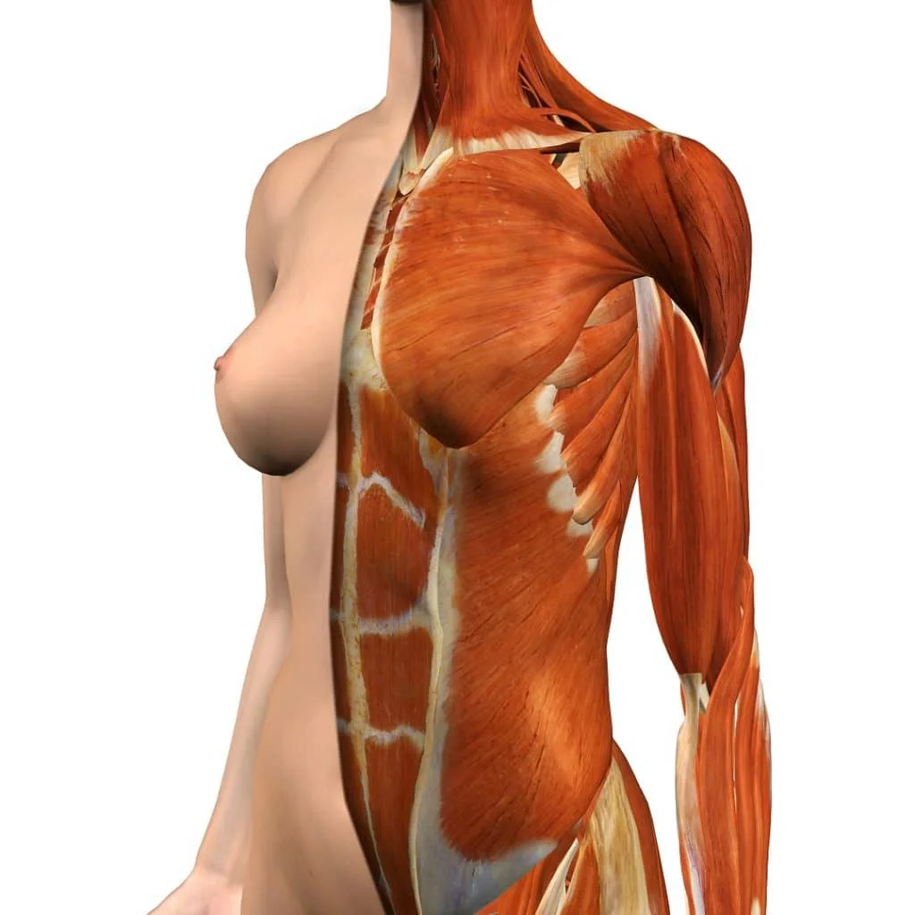
The uppermost or superficial layer of your abdomen contains the skin, subcutaneous tissue, two layers of fascia (Scarpa’s fascia and Camper’s fascia), and also connective tissue. This part of the abdomen gives structure and protection to your internal organs. Nerves, lymphatics, and blood vessels are also present throughout.
Abdominal Skin
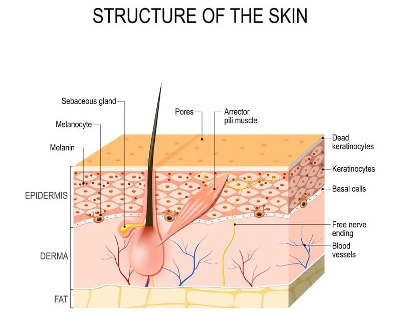
Skin quality is an important indicator of wellness. Strong abdominal skin is firm and elastic. Unfortunately, the older you get, the easier it is to damage the skin because senior skin does not bounce back as easily as it once did.
Gaining weight or having a baby can also take a toll on skin. As the body stretches to accommodate either underlying fat or a growing fetus, it can weaken the skin structure dramatically. The skin may develop a loose appearance, with creases and folds. Stretch marks are another common problem.
Stretched skin does not always shrink back to how it was before. Women with multiple pregnancies and patients who have undergone bariatric or gastric bypass surgery are most at risk. People over age 30 are also affected because skin produces progressively less collagen and elastin as the body ages.
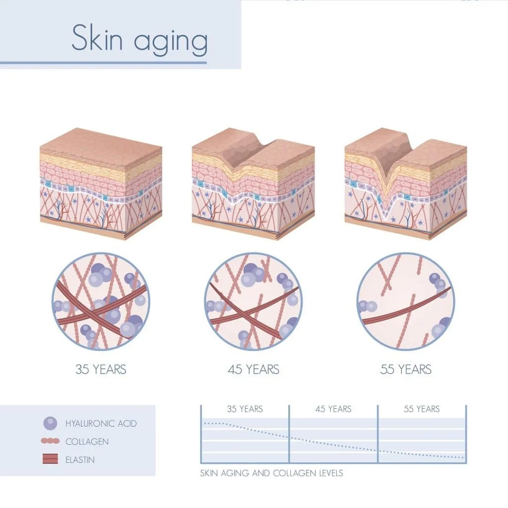
There are many ways to treat skin ageing and stretch marks. Lots of over-the-counter beauty products, home remedies, and prescriptions promise to make the skin appear new again. It is important to mention that cosmetic surgery does not change the biology of the skin. If your skin is aged, inelastic, or lacking collagen, an abdominoplasty won’t make your skin inherently new again. No surgeon can promise that with body contouring surgery.
However, abdominoplasty can help your skin appear fresher since the skin is pulled nice and taut when the incision is closed with sutures. This gives the skin a firmer appearance, even though the skin is not structurally different than it was before surgery. Abdominoplasty may also remove some stretch marks when the skin is trimmed.
Subcutaneous Tissue
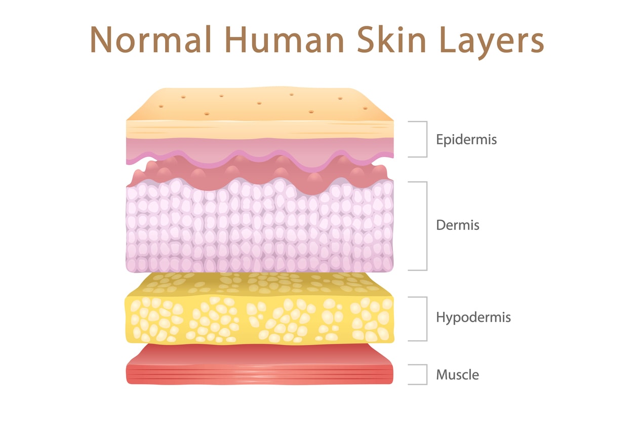
The layer just below the skin is called subcutaneous tissue. It is also known as the hypodermis. From regulating body temperature to blood flow, this tissue serves many important functions. It is made up of fat and connective tissue. Blood vessels and nerves run through this layer, which acts as a passageway for blood flow between the upper layers of skin and underlying muscle.
Camper’s Fascia
Below the skin are two layers of superficial fascia. The Camper’s fascia is the first layer. The fascia of Camper contains mostly areolar tissue, which is made of elastic and smooth connective fibers with a minimal amount of fat.
Areolar Tissue
There are six types of connective tissue in the human body. Areolar tissue is a loose connective tissue made from a meshwork of collagen fibers and elastic tissue. One function of areolar tissue is to connect the skin to the muscles below. Areolar tissue helps give the abdomen structure so that everything stays in its proper place. It also stores some fat and helps insulate the body.
Adipose Tissue (fat)
Fat, or adipose tissue, is a special kind of connective tissue made of adipocytes. The purpose of these cells is to store energy from food. Food fuels adipose tissue with calories, which gives us energy. That’s why when you workout, you burn fat. Yet even when you sit still or sleep, your body is always burning calories. Whether you are digesting food, breathing, or blinking your eyes, the human body is always doing something that requires energy. Adipose tissue also cushions and insulates the body.
Too much subcutaneous fat can leave your abdomen soft and pudgy. When we think of getting surgery performed, it’s often this layer of connective tissue that we want to focus on. Pregnancy, ageing, medications, eating too many high-calorie foods without working out, and even your body’s unique biochemistry can lead to a buildup of adipose tissue.
The mid-section is often the first place people gain weight. Lipo-abdominoplasty surgery is an excellent way to remove belly fat, even the kind that exercise and diet alone don’t seem to help.
Scarpa Fascia
The Scarpa fascia is located beneath the Camper’s fascia in your abdomen. It is a thick, membraneous layer on the anterior abdominal wall.
The Scarpa fascia is a very important part of successful abdominoplasty surgery. This is the structure that gets sutured closed in abdominoplasty. It is very strong and tough, so it can support a lot of tension. That allows your surgeon to get a nice, taut closure of the incision without compromising skin vascularity.
Abdominal and Core Muscles
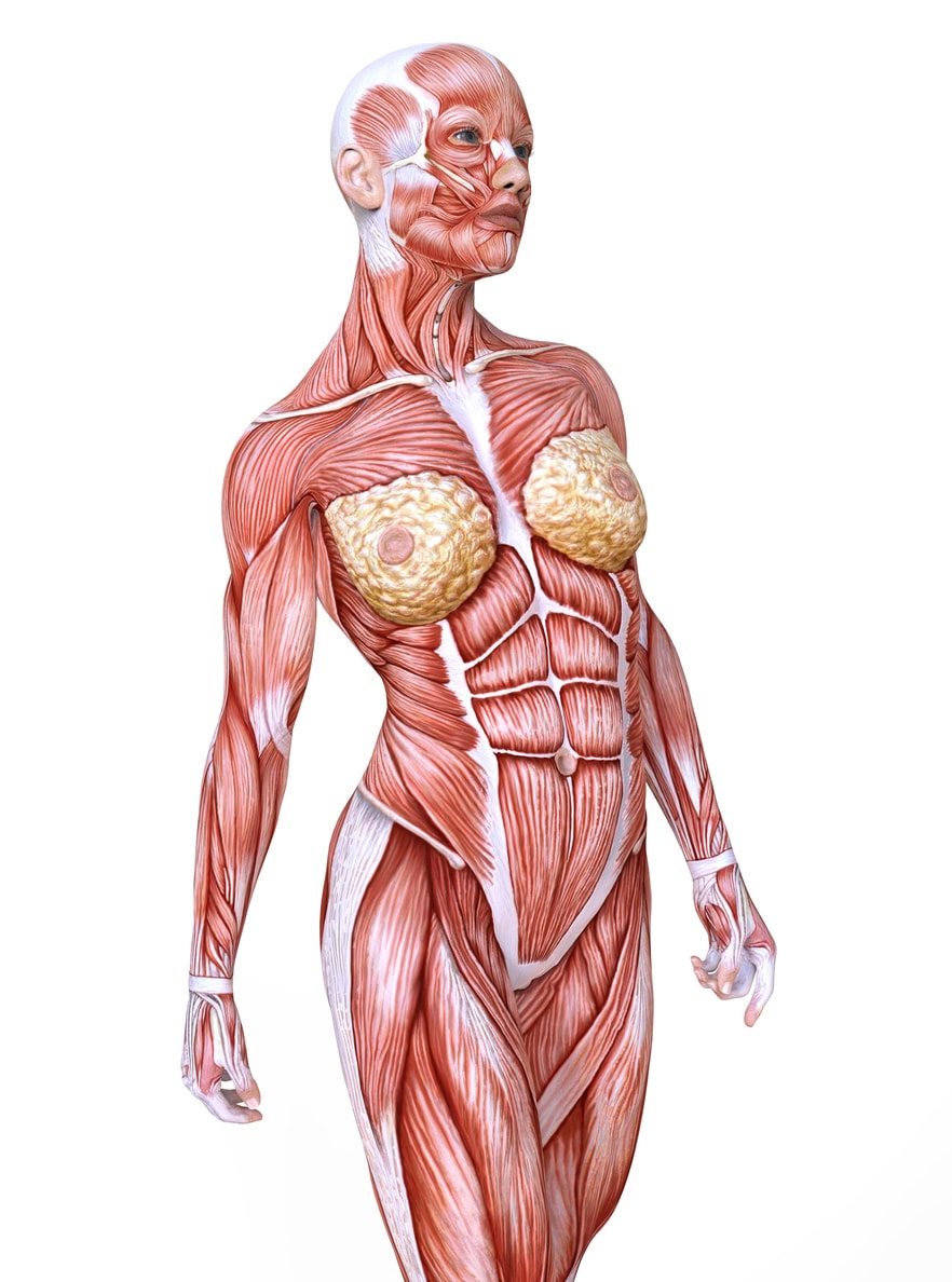
Your body core includes all of the muscles located in your midsection, including those found on the front, back, and sides. These muscles work together to give your body proper alignment, mobility, strength, and the ability to bear weight. The abdominal and pelvic floor muscles work in tandem to keep you pain-free. A weak body core can cause all sorts of problems, including pain.
Inside your abdomen are some very important muscle groups.
Rectus Abdominis Muscles
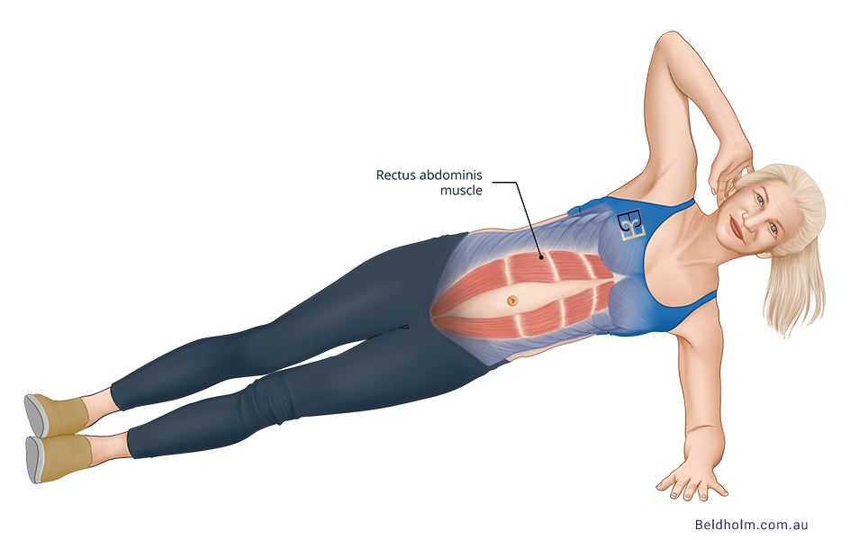
The most commonly known abdominal muscle group is the rectus abdominis. These are known as the “six-pack” muscles. This is the most superficial abdominal muscle group because it lies closest to your skin. You can see these muscles very clearly in a person with a muscular physique.
The rectus abdominis is a flat muscle made of layers that form a fibrous sheath. The fibrous bands that divide the abs into the small, rectangular sections that you see on a toned tummy are called tendinous intersections.
The rectus abdominis is also divided down the middle of your abdomen. This gives the rectus the appearance of having two sides, located vertically on either side of your belly button. A tough, fibrous structure known as the linea alba joins them at the midline. The linea alba is rather elastic, and for good reason. It helps support the internal abdominal stress that is created when you move.(ref 3) Whether you are lifting, pulling, twisting, or bending, the linea alba adjusts to accommodate your body’s many incredible movements using a network of elastic fibers.
The linea alba also stretches during pregnancy. As the baby grows, the tummy enlarges and the linea alba must stretch to make room for the growing baby. Unfortunately, it can only stretch so much before it tears. And once it stretches, it may not go back to how it was before pregnancy.
Known as diastasis recti, abdominal muscle separation is a condition that is very common among pregnant women. It can cause belly bulge, back and pelvic pain, urine leaking, and a visible gap between the rectus muscles on the tummy. Abdominal muscle separation is a leading reason women get body contouring surgery after giving birth. Straining, poor posture, and lifting heavy weights can also stretch or tear the rectus and connecting membrane.
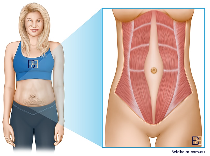
The muscles do not always go back to how they were before pregnancy. Core exercises can help prevent and treat muscle separation, but for some patients the damage may be beyond the help of exercise alone. Abdominoplasty can repair diastasis recti caused by pregnancy. During surgery, Dr. Beldholm uses sutures to bring the muscles back to their original position.
External Obliques Muscles
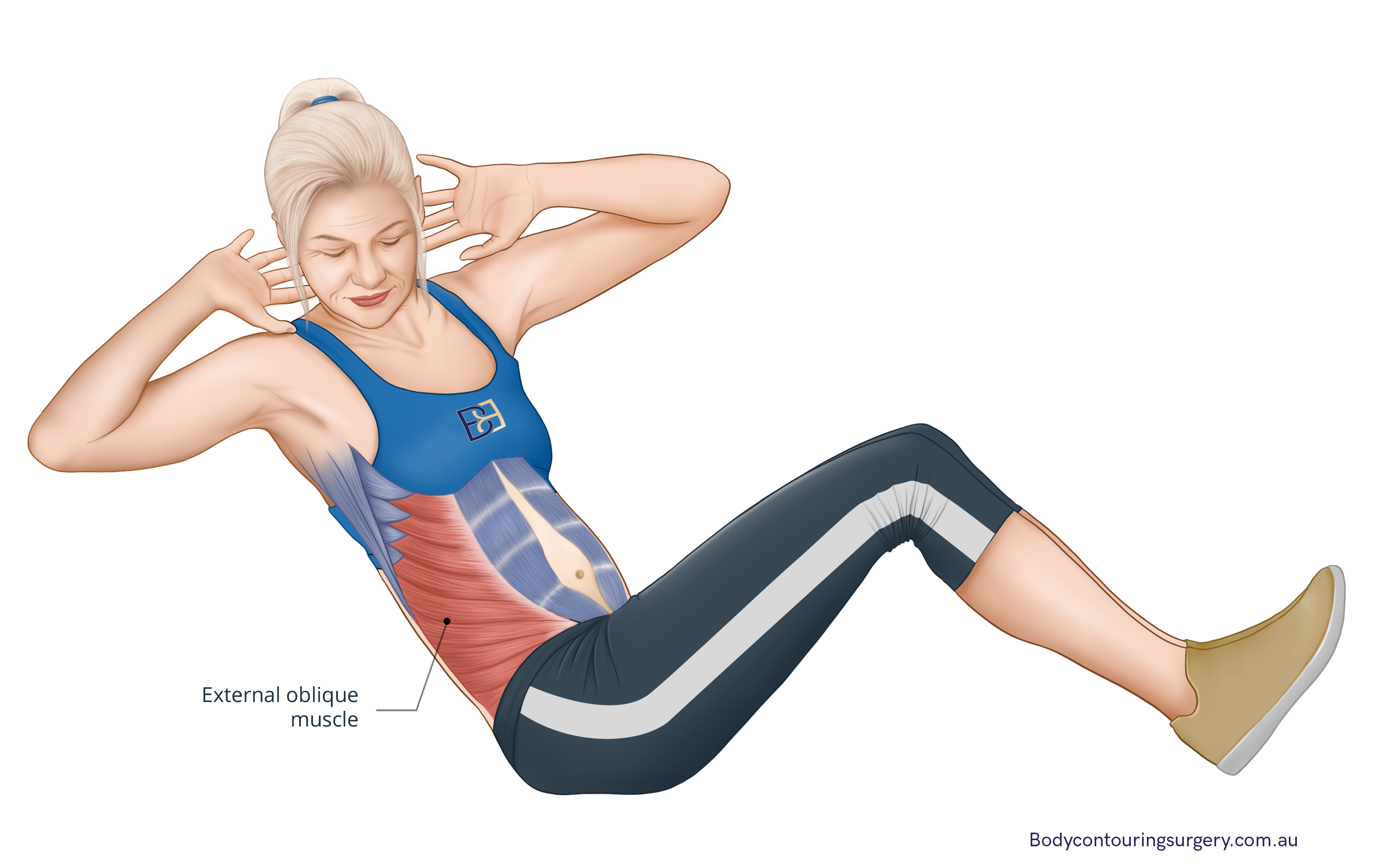
The external oblique muscles are located superficially on either side of your six pack muscles. They run along the sides of your abdomen. Twisting, turning, and bending engage the external obliques. These muscles work in tandem with all your other abdominal muscles to give your abdomen strength. Strong external obliques allow you to live an active lifestyle. The external obliques attach to the ribs.
Internal Obliques Muscles
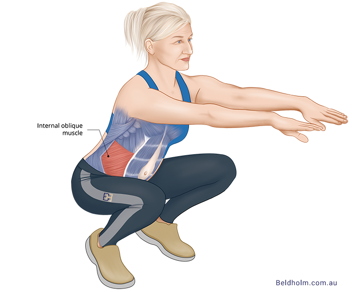
The internal obliques have a similar function to the external obliques. They are located deeper in the abdomen. They attach to the ribs and linea alba. Both the internal and external oblique muscles rely on each other to flex and rotate your torso. Internal obliques are sometimes called “same side rotators”. Like the external obliques, they aid in supporting your abdominal wall.
Transversus Abdominis Muscles

Your deepest abdominal muscles are known as the transversus abdominis. It is located below all your other abdominal muscles and above your rib cage. When you perform core body exercises, these are the main muscles that you are working out.
The transversus abdominis is vital for good posture and lumbar support. It also helps hold the abdominal organs in place. If you have diastasis recti from pregnancy, this is the group of abdominal muscles you should focus on exercising. Doing so can reduce back pain from diastasis by increasing spinal support. (ref 4)
When you have diastasis recti from pregnancy, regaining core strength is essential. The transversus abdominis are the most important muscle group to work out in early recovery. This deep muscle group sets the foundation for all your other abdominals to work effectively. A fine-tuned body core keeps your core strong.
Pyramidalis Muscle
The pyramidalis muscle is a small, triangular muscle located in front of the rectus abdominis. Its function is to tense the linea alba, which helps maintain the integrity of the abdominal wall. Interestingly, this muscle is not present in all individuals, with about 20% of people lacking it entirely.
Despite its variability, the pyramidalis muscle plays a supportive role in the overall structure of the abdominal wall.
Structures Below the Core Muscles
The transversal fascia and peritoneum are both protective membranes that help keep your inner organs intact. They aid in supporting your organs, giving your abdomen yet another layer of protection and strength.
Transversalis Fascia
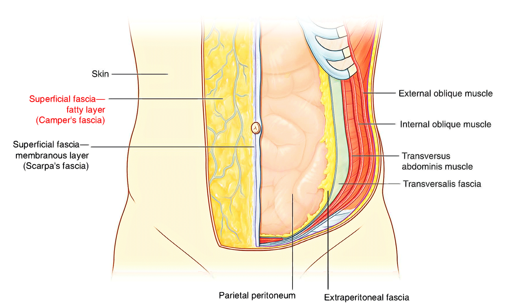
The transverse fascia is located between the transversus muscle and the peritoneum. It lines the anterolateral abdominal wall. It is thick and dense, made up of tough fibers. In patients with hernia, a weak transversalis fascia is usually the culprit. When this fascia is damaged or weakened, the intestines can bulge through, causing pain when you bend, cough, or lift a heavy object. Hernias can be pretty painful.
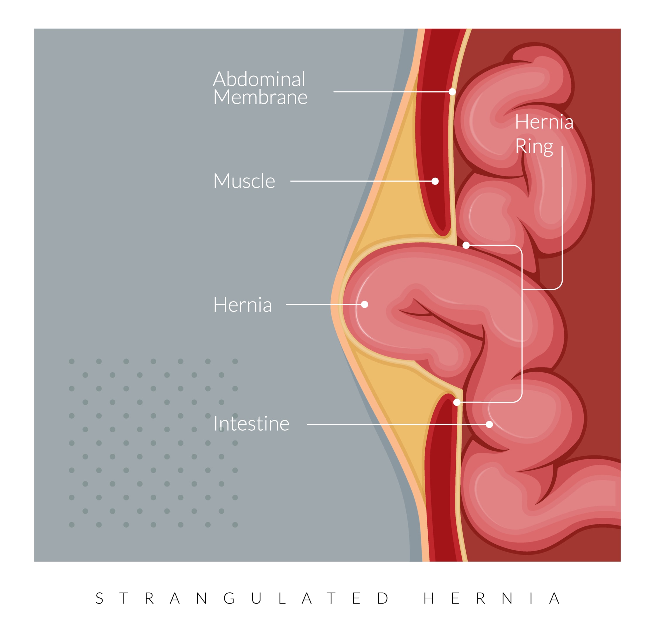
Increased uterine pressure during pregnancy can also sometimes cause umbilical hernias. You may notice swelling or a bulge near the navel. Umbilical hernias are not dangerous per se, and they won’t harm the baby, but they can cause discomfort. Surgery is the recommended treatment for hernias because they do not go away on their own.
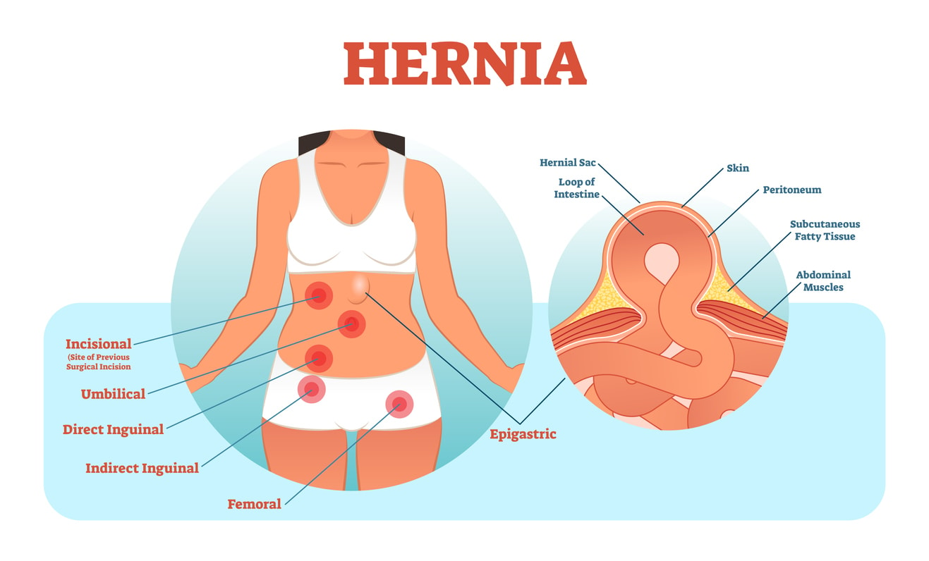
Having a strong transversalis fascia can reduce your risk of hernias. But the transversalis fascia is a membrane, not a muscle. So there is not much you can do to regain strength to the membrane. However, it is always a good idea to eat healthy foods and maintain a proper body weight to help keep all parts of your body working properly.
Peritoneum
The peritoneum is a serous membrane that lines the abdominal cavity and encases all your organs (except for the adrenal glands and kidneys). It is an important part of your abdominal wall anatomy. Not only does it give support to your abdominal organs, it also acts as a passageway for nerves, blood vessels, and lymphatics.
Blood Supply and Innervation of the Abdominal Wall
The blood supply and innervation of the abdominal wall are crucial for its health and functionality. Proper blood flow ensures that the abdominal muscles and tissues receive the necessary oxygen and nutrients, while the nerve supply facilitates movement and sensation.
Understanding these aspects is essential for both medical professionals and patients undergoing abdominal procedures.
Arterial Supply
The arterial supply to the abdominal wall is vital for its maintenance and healing. The major arteries include the superior epigastric artery, which originates from the internal thoracic artery, and the inferior epigastric artery, which arises from the external iliac artery. Together, these arteries ensure a consistent blood supply to the abdominal wall, supporting its various structures and functions.
Proper arterial supply is crucial for the health and functionality of the abdominal wall, providing oxygenated blood essential for sustaining the muscles and other tissues. Understanding the arterial network is vital for surgical planning and recovery.
Venous Drainage
The venous drainage system of the abdominal wall mirrors the arterial supply, with veins accompanying the arteries and following similar paths. The inferior epigastric vein, a key vessel in this system, drains into the external iliac vein, playing a significant role in the deep venous drainage of the abdomen.
Efficient venous drainage is essential for removing deoxygenated blood and waste products from the abdominal tissues.
Nervous Supply
The nervous supply to the anterolateral abdominal wall is primarily from the T7 to T12 intercostal nerves, which provide both sensory and motor innervation. These nerves are crucial for the functionality and sensation of the abdominal wall, facilitating movements and responding to stimuli.
In addition to the thoracoabdominal nerves, the iliohypogastric and ilioinguinal nerves from the lumbar plexus are essential for innervating the internal oblique and transversus abdominis muscles. Recognizing the nerve supply is crucial for surgical procedures and effective recovery, ensuring optimal muscle function without complications.
Hernias
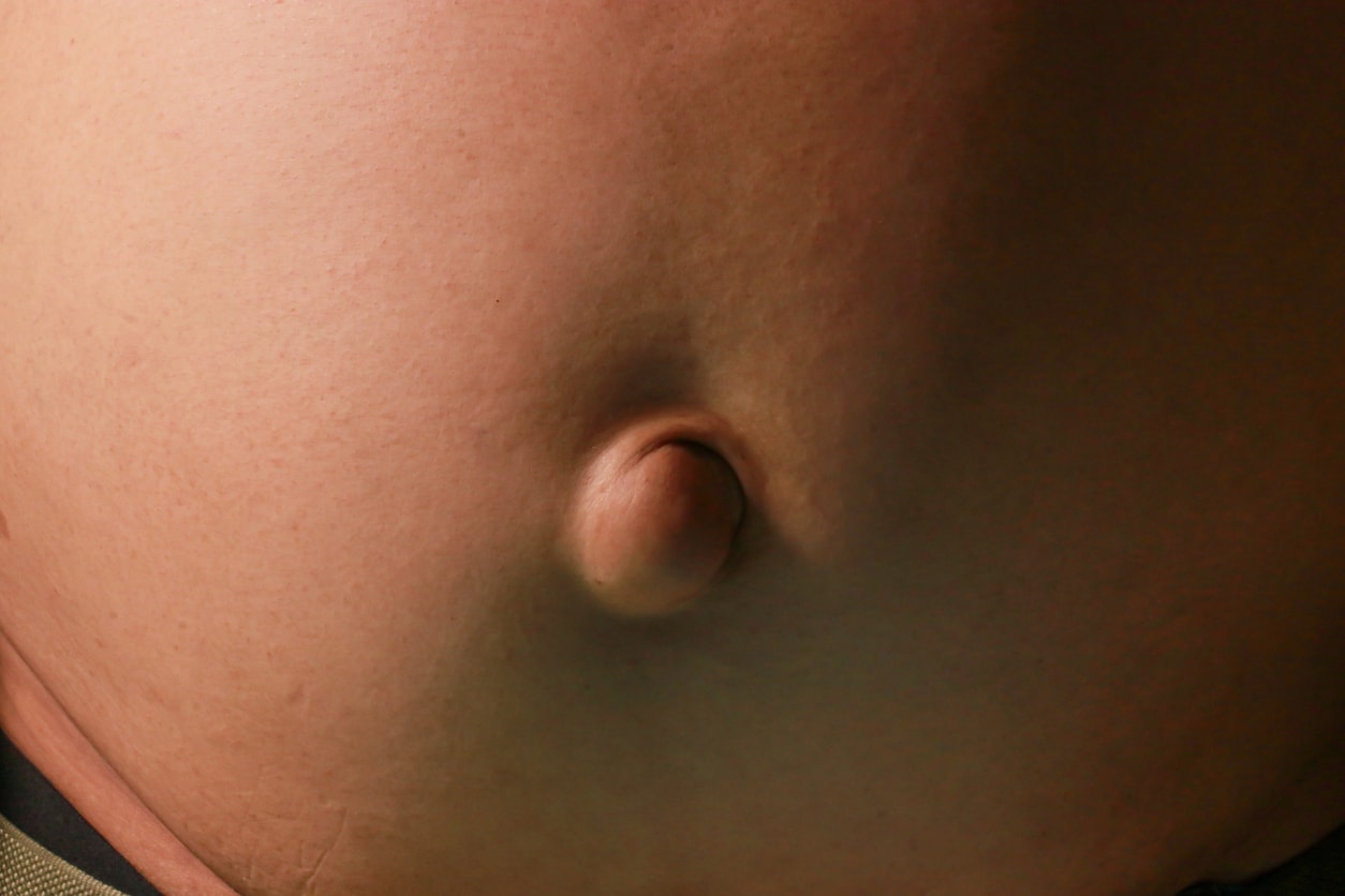
Hernias occur when an internal organ pushes through a weak spot in the abdominal muscles, creating a noticeable bulge. Different types of hernias, such as umbilical and inguinal hernias, can arise due to congenital weaknesses or acquired factors like heavy lifting and chronic coughing. Understanding the anatomy of the abdominal wall helps in diagnosing and treating these conditions effectively.
For instance, umbilical hernias involve the herniation of abdominal contents through a patent umbilical ring, which can often resolve spontaneously. In contrast, Spigelian hernias occur at the semilunar line and are more complex, requiring surgical intervention for proper management. Recognizing these variations is crucial for effective treatment and patient recovery.
Dr Beldholm’s Final Conclusions

As a specialist surgeon, I can attest to the complexity and vital importance of the abdominal wall. This intricate structure not only protects our internal organs but also facilitates movement and maintains core stability, which are essential for living an active lifestyle.
Understanding the detailed anatomy of the abdominal wall is crucial, whether you are considering surgical procedures like abdominoplasty or aiming to rebuild your core strength through exercise. A comprehensive grasp of this anatomy allows for better surgical outcomes and more effective rehabilitation.
In conclusion, whether you are a patient or a medical professional, recognising the significance of the abdominal wall’s anatomy is key to achieving optimal wellness and successful outcomes.
Frequently Asked Questions
Why is understanding the abdominal wall important for abdominoplasty?
Understanding the abdominal wall is crucial for abdominoplasty because it helps set realistic expectations and tailors the surgical technique to your unique anatomy, leading to better results.
What are the main layers of the abdominal wall?
The main layers of the abdominal wall are skin, superficial fascia, muscular layers, and deep fascia. Each of these layers is important for protection and support of the abdomen.
How do muscle strains in the abdominal wall occur?
Muscle strains in the abdominal wall usually happen due to heavy lifting, sports, or even intense coughing and sneezing, which can cause tiny tears or pulls in the muscles. So, be careful when pushing your limits!
What is diastasis recti, and how is it treated?
Diastasis recti is when the abdominal muscles separate, usually during pregnancy. Treatment often involves physical therapy, and in more severe instances, surgery might be needed.
What exercises are recommended for core stability?
To build up your core stability, try modified planks and single-leg abdominal presses.
Book your appointment online now
References
- Farage, Miranda A., et al. “Characteristics of the Aging Skin.” Aging Clinical and Experimental Research, vol. 20, no. 3, 2008, pp. 195–200., doi:10.1007/bf03324769.
- Hunstad, Joseph P., and Remus Repta. Atlas of Abdominoplasty. Saunders/Elsevier, 2009.
- Pulei, A N, et al. “Distribution of Elastic Fibres in the Human Abdominal Linea Alba.” Anatomy Journal of Africa, vol. 4, no. 1, 2015, ajol.info/index.php/aja/article/view/118732.
- Sancho, M.f., et al. “Abdominal Exercises Affect Inter-Rectus Distance in Postpartum Women: a Two-Dimensional Ultrasound Study.” Physiotherapy, vol. 101, no. 3, 6 May 2015, pp. 286–291., doi:10.1016/j.physio.2015.04.004.
- Varani, James, et al. “Decreased Collagen Production in Chronologically Aged Skin.” The American Journal of Pathology, vol. 168, no. 6, 2006, pp. 1861–1868., doi:10.2353/ajpath.2006.051302.

