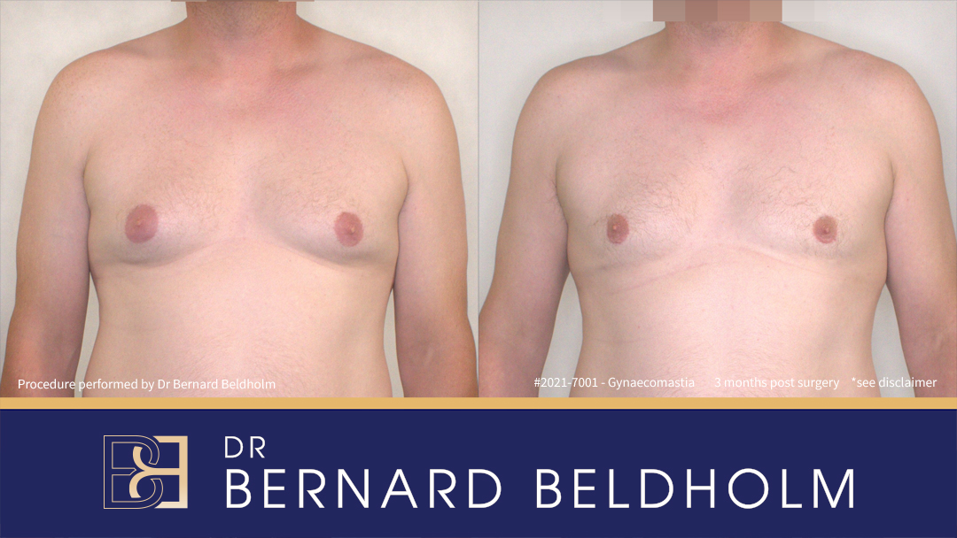
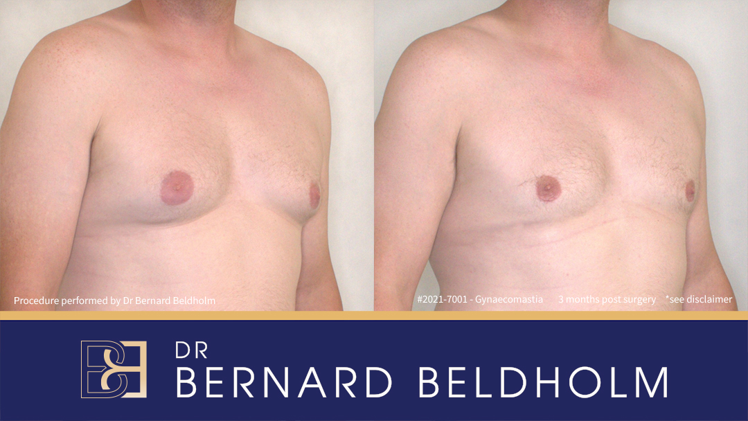
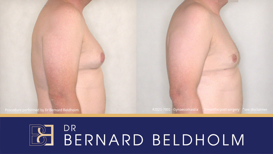
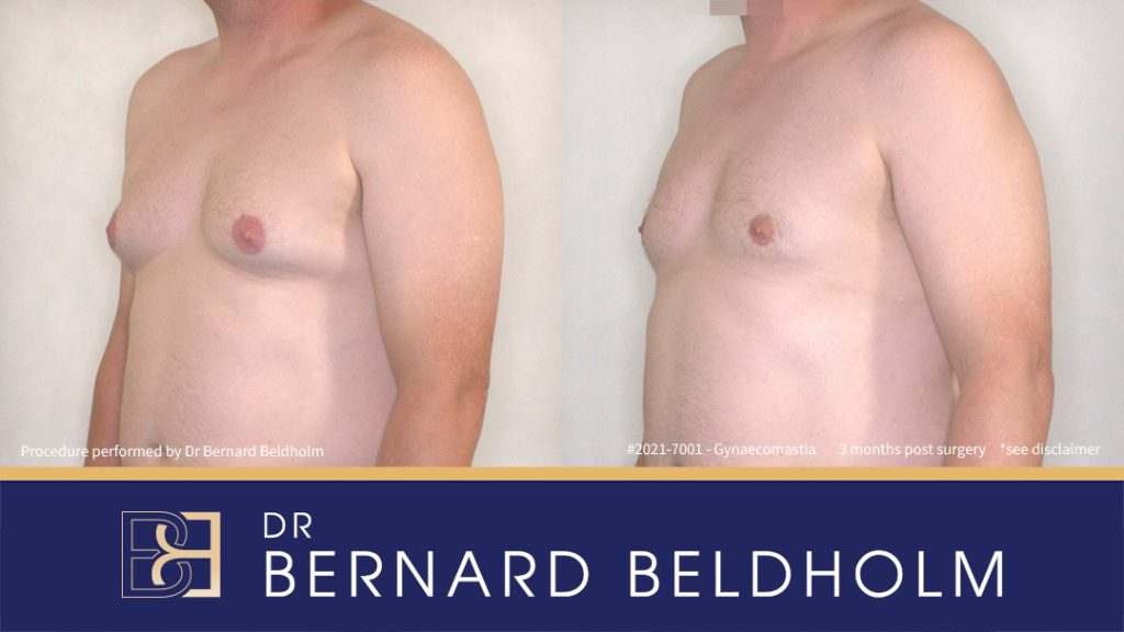
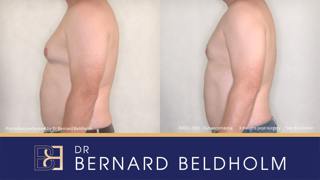
*Results vary
Patient History
- Male patient, 25 years old.
- Longstanding history of gynaecomastia (benign enlargement of male breast tissue), first noted during adolescence.
- Clinical examination identified:
- Fibroglandular tissue beneath the nipple-areolar complex (NAC).
- A larger component of pseudo-gynaecomastia (adipose tissue hypertrophy of the chest wall).
Classification
- Condition classified using Dr Beldholm’s internal grading system: Grade BB 2b.
- Recommended management: ultrasound-assisted suction lipectomy (VASER liposuction).
- Due to the presence of a small amount of fibroglandular breast tissue beneath the areola, sub-areolar excision (direct glandular excision) was also performed.
Operation Performed
- Bilateral ultrasound-assisted suction lipectomy (VASER liposuction) performed on the anterior chest and lateral chest wall.
- Peri-areolar incisions were made bilaterally to allow removal of glandular breast tissue.
- A 10 French Belovac drain was placed on each side, removed the following day.
Histopathology Report
Macroscopic findings
- Right breast specimen: Fibroadipose breast tissue, 9g, 77×25×15mm. Cut surface: uniform fibrofatty tissue. No discrete macroscopic lesions.
- Left breast specimen: Fibroadipose breast tissue, 10g, 64×35×12mm. Cut surface: uniform fibrofatty tissue. No discrete macroscopic lesions.
Microscopic findings
- Right breast specimen: Fibrofatty breast tissue with occasional ducts showing columnar cell change without cytological atypia. Apocrine metaplasia present. No in situ or invasive malignancy. No microcalcifications.
- Left breast specimen: Fibrofatty breast tissue with unremarkable ducts and densely fibrous stroma. Features may represent late-stage gynaecomastia. No in situ or invasive malignancy. No microcalcifications.
Final Diagnosis
- Findings consistent with gynaecomastia (benign enlargement of male breast tissue).
Images:
3 months after the operation
All procedures have potential complications. For more information see: Disclaimer – Dr Bernard Beldholm Specialist Surgeon
Download our free e-book: Gynaecomastia Surgery – Must Know Facts for Men
Click HereThis gallery shows gynaecomastia surgery before and after videos and photos.
Disclaimer: The content on this website is considered Adult content. Individual results may vary. All surgery carries risks. You should seek a second opinion before proceeding. The opinions that are expressed on this website are those of Dr Bernard Beldholm & these opinions may differ from other doctors’ opinions.
The information provided on and through this website is not medical advice and should not be relied on. It is “best efforts” and for general information only. Do not use this website as a substitute for medical advice or self or other diagnosis. Dr Bernard Beldholm & Body Contouring Surgical Clinic Pty Ltd accepts no liability for any error, omission, use of or reliance on the materials provided on the website.
Dr Bernard Beldholm (MED0001186274) M.B.B.S B.Sc (Med) FRACS, is a Registered medical practitioner, Specialist surgeon (specialist registration in Surgery – general surgery). See full Disclaimer

